
APA Style
Abhishek Vijukumar, Kamaljeet, Harkomal Singh, Tarun Kalra, Bintoo Sharma. (2025). A Century of Alkaptonuria: Evolving Insights into a Rare Metabolic Disorder. GenoMed Connect, 2 (Article ID: 0019). https://doi.org/10.69709/GenomC.2025.122212MLA Style
Abhishek Vijukumar, Kamaljeet, Harkomal Singh, Tarun Kalra, Bintoo Sharma. "A Century of Alkaptonuria: Evolving Insights into a Rare Metabolic Disorder". GenoMed Connect, vol. 2, 2025, Article ID: 0019, https://doi.org/10.69709/GenomC.2025.122212.Chicago Style
Abhishek Vijukumar, Kamaljeet, Harkomal Singh, Tarun Kalra, Bintoo Sharma. 2025. "A Century of Alkaptonuria: Evolving Insights into a Rare Metabolic Disorder." GenoMed Connect 2 (2025): 0019. https://doi.org/10.69709/GenomC.2025.122212.
ACCESS
Review Article
Volume 2, Article ID: 2025.0019

Abhishek Vijukumar
2216730@isfcp.org
Kamaljeet
2116957@isfcp.org
Harkomal Singh
harkomalsingh1993@gmail.com
Tarun Kalra
tarunkalra222@gmail.com
Bintoo Sharma
bintoo1053ps.phd25@chitkara.edu.in
1 Department of Pharmacy Practice, ISF College of Pharmacy, Moga 142001, Punjab, India
2 Department of Pharmacy Practice, Chitkara College of Pharmacy, Chitkara University, Rajpura 140401, Punjab, India
* Author to whom correspondence should be addressed
Received: 25 Jul 2025 Accepted: 22 Dec 2025 Available Online: 23 Dec 2025 Published: 29 Dec 2025
Alkaptonuria is a rare autosomal recessive disorder caused by a deficiency of the enzyme homogentisate 1,2-dioxygenase (HGD). When HGD does not function properly, homogentisic acid (HGA) accumulates in the body despite the kidneys’ capacity to excrete it regularly. The diagnosis of AKU may not occur until the ochronotic arthropathy manifests, which usually happens in adulthood. There is a paucity of information on the condition’s early stages and infancy. The formation of darkly pigmented urine, dried urine spot (DUS) analysis, immunofluorescence, Thioflavin T (ThT) staining, Congo Red (CR) staining, and transmission electron microscopy (TEM) may all be used to detect this condition. Management strategies include ascorbic acid supplementation, dietary restriction of phenylalanine and tyrosine, and pharmacological intervention. Nitisinone is the only medication approved by the European Medicines Agency as a disease-modifying treatment for individuals with alkaptonuria and has demonstrated significant therapeutic potential.
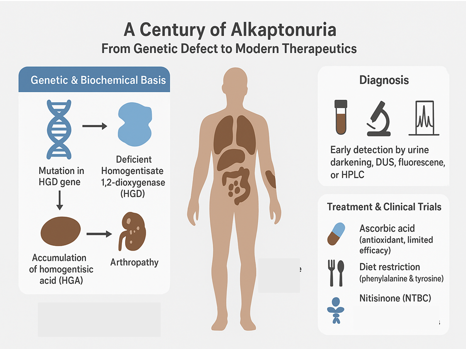
In 1908, Sir Archibald Edward Garrod identified alkaptonuria (AKU) as one of the four recognized congenital metabolic disorders [1]. In addition, this was the first disease to be classified as matching Mendelian recessive inheritance. There is a historical background to the illness, with many reports describing people displaying symptoms of AKU. In 1584, Scribonius reported a case of a young boy whose urine resembled ink. Research indicates that alkaptonuria (AKU) affects approximately 1 to 8 per 1,000,000 newborns [2]. 1 in 19,000 is the highest rate found in the Dominican Republic and in the Piestany area in Slovakia [3]. Alkaptonuria (AKU) is a rare autosomal recessive disorder caused by a deficiency of the enzyme homogentisate 1,2-dioxygenase (HGD). In 1993, genetic research on families of Slovak patients identified the HGD enzyme gene at 3q21–q23 on chromosome 3 [4]. Homogentisate 1,2-dioxygenase (HGD) is an enzyme that catalyzes the conversion of homogentisic acid (HGA) into maleylacetoacetic acid during the catabolism of phenylalanine and tyrosine. Biochemically, HGD dysfunction causes HGA to remain in the body, despite the kidneys removing it at their normal rate [5]. Large amounts of homogentisic acid (HGA) are oxidized via a benzoquinone acetate intermediate, resulting in the formation of pigmented polymers. In ochronosis, HGA pigment accumulates in several tissues, mainly in connective tissues, joints, and spinal cartilage, tendons, and ligaments. Due to increased weakness and susceptibility, the connective tissues in ochronosis allow decay to occur, which may result in discoloration of the ears, eyes, and skin, as well as the formation of calculi in the kidneys, prostate, gallbladder, and salivary glands [6]. In many cases, individuals with this condition experience renal failure, ruptures of tendons, ligaments, and muscles, fractures, reduced bone density (osteopenia), and spinal disorders that compromise stability (spondyloarthropathy). They may also develop abnormal spinal curvatures, including kyphosis and scoliosis, and often require joint replacement surgery [7], as illustrated in Figure 1. Therefore, several degenerative processes, inflammation, and calcification may develop, causing the disorder to progress into severe spondyloarthropathy and osteoarthritis. Research has shown that X-ray changes in the joints typically begin around the age of 30, with the first joint replacement most commonly occurring at approximately 55 years of age [8]. At the age of 54, heart valve issues are common, whereas coronary artery disease is seen most often at 59 years of age. Early diagnosis of the condition is challenging because only a few cases are identified in infancy. Consequently, the disorder is frequently not diagnosed until the child begins to experience back, joint, and muscle pain. Prior to 2011, treatment for AKU was limited and involved only pain-relieving drugs and arthroplasty [8,9]. Studies where vitamin C acts as an antioxidant lowered the amount of benzoquinone acetic acid, but had no impact on HGA levels [10]. A low-protein diet is recommended and may provide therapeutic benefits; however, adherence to such a diet is often challenging. A study has reported a treatment using the drug nitisinone (NTBC), which inhibits 4-hydroxyphenylpyruvate dioxygenase, and demonstrated its efficacy in managing the disease [11].
The HGD gene, located on chromosome 3q21–q23, encodes the homogentisate 1,2-dioxygenase (HGD) enzyme. Mutations in this gene lead to loss of enzyme function, contributing to the clinical manifestations of alkaptonuria (AKU) [12]. There has been significant research into the genetics of AKU, and many types of changes in the HGD gene have been found in patients worldwide. They consist of missense, nonsense, splicing, small deletion/insertion, and large-scale rearrangement mutations. Identifying and describing HGD mutations in AKU has helped in better understanding the genetic causes of AKU symptoms. Several changes in the HGD gene can cause diverse levels of enzyme deficiency, which lead to a wide range of symptoms, from acute to chronic forms of the disease [13]. As a result, genetic testing and counseling programs are now offered to those with AKU and their family members through molecular-level research. The frequency of AKU varies among different ethnic groups, with increased incidence rates seen in some populations, such as the Dominican Republic and Slovakia. Founder mutations and mutation hotspots have been identified in these populations, helping to explain the increased incidence of alkaptonuria (AKU) in certain regions [14]. AKU has also been studied in Slovakia due to its greater prevalence than in other nations. In Slovakia, researchers have studied both the genetic details of HGD mutations and the medical and biochemical features of AKU among the population. Through this research, scientists have obtained valuable insights into the genetic background of alkaptonuria (AKU) in Slovakia and have generated distinctive data for genotype-phenotype comparisons within this population [15]. Understanding the molecular aspects of AKU can reveal novel treatment opportunities. It is vital to understand the HGD mutations in AKU patients to develop targeted molecular treatments, such as gene therapy or small-molecule interventions aimed at repairing the HGD enzyme. This area of research may lead to hopeful treatments for AKU in the future. Overall, HGD mutations have been identified and studied, enabling correlations between genotype and disease, revealing population-specific variations in alkaptonuria mutations, and providing prospects for the development of new therapeutic approaches [16]. Researchers are making steady progress toward understanding AKU and identifying suitable therapies for this uncommon metabolic disorder.
Diagnosis may take time, since the arthropathy usually develops when someone reaches adulthood, and the earliest details of AKU are not widely known [17]. The delay in the appearance of dark urine depends on urine acidity, and such delays may lead to misdiagnosis as another condition [18]. There is no clear connection between the amount of HGA in the blood and symptoms, age, or sex [8,9]. This can be diagnosed using dried urine spot (DUS), Congo Red (CR) staining, Thioflavin T (Th-T) staining, immunofluorescence, and transmission electron microscopy (TEM), as shown in Figure 2. Dried urine spots (DUS) are used to detect hereditary disorders, including sporadic cases of alkaptonuria (AKU), primarily as a qualitative diagnostic tool. A dark brown hue is widely known to emerge when alkali (e.g., sodium hydroxide) is added to either HGA solutions or AKU urines [18]. Early detection of AKU would help researchers better understand the disease and relate it to physiological processes. In alkaptonuria (AKU), homogentisic acid (HGA), a reducing agent containing two phenolic groups, is converted into benzoquinone acetic acid, which then acts as an oxidizing agent to produce a pigment resembling melanin [9]. Identifying secondary amyloidosis in AKU may be difficult, but it is critical to rapidly recognize and treat the condition. According to a 2014 study by Millucci et al., published in Diagnostic Pathology, selecting relevant specimens is critical for accurate diagnosis. The researchers observed that amyloid was detected in only one specimen from the abdominal fat pad, although all patients had amyloid accumulation in salivary glands and in other organ biopsies. This shows that salivary glands are the best choice for detecting amyloid in the early stages of AKU. The authors discussed the importance of performing Congo Red (CR) staining, Thioflavin T (Th-T) staining, immunofluorescence, and TEM to detect amyloid deposits in different parts of the body [19]. Individuals with alkaptonuria (AKU) exhibit considerable variability in their clinical manifestations. The study gives a detailed outline of the patients involved in the examination. The features involved cover peripheral neuropathy, orthostatic hypotension, enlarged ventricles and atria, chronic bacterial infection, spondyloarthropathy, swollen tongue, and surgeries in the past [20]. SAA, HGA in plasma and urine, and amyloid plaque occurrence were checked for each patient. Assessments were conducted to identify which specimens are most useful for the rapid detection of amyloid in AKU patients. Gabriella Jacomelli’s report, titled "Quick Diagnosis of AKU by Homogentisic Acid," was used in this study [21]. Two approaches for easy AKU testing using HGA measured in dried urine spots are the main focus of the report titled “Determination in Urine Paper Spots.” In the first method, alkaline conditions cause the color to appear quickly and reliably, and allow for some numerical measurements. The second method uses the sensitive, quantitative approach of HPLC (High Performance Liquid Chromatography). With these approaches, AKU patients can rely on accurate screening [21-23].
As stated in Figure 3, the therapies that have been examined for AKU include: 4.1. Tyrosine and Phenylalanine-Restricted Diet A protein-restricted diet has been recommended as a viable way to minimize HGA production. This therapeutic strategy has shown success in treating numerous genetic metabolic illnesses and has exhibited good effects on neurological outcomes and overall survival [24]. 4.2. Vitamin C (Ascorbic Acid) Ascorbic acid has been examined for its effects on alkaptonuria, particularly its potential to block the binding of homogentisic acid in connective tissue [25]. Studies have also explored the effects of ascorbic acid on HGA excretion in urine. 4.3. Nitisinone Nitisinone has shown promise as a specific treatment for alkaptonuria. It has been tested in people with AKU and has been associated with positive effects, including the suppression of ochronotic osteoarthropathy [26]. Nitisinone works by reducing HGA production through the enzyme HPPD. Reducing this enzyme’s activity with nitisinone lowers HGA levels, helping combat ochronosis and its associated symptoms. According to studies, including SONIA 2, nitisinone can reduce HGA levels by nearly 100 percent and improve symptoms in people with AKU. Since HGA levels have dropped, the disease progresses more slowly, as seen by the slower increase in the AKU Severity Score Index (AKUSSI) [23]. The European Medicines Agency recently authorized nitisinone as the first therapy to slow the progression of AKU in patients. Research has determined that nitisinone works to prevent ochronosis in mouse models of AKU and can also slow the development of the disease when given to human patients with clear signs of AKU [27].
5.1. SONIA 1 A major research study, the Suitability of Nitisinone in AKU (SONIA 1), is examining the effectiveness and safety of nitisinone for patients with AKU. The researchers tested different doses of nitisinone in two groups to assess its impact on urinary HGA excretion. In the SONIA 1 trial, 40 patients with AKU were randomly assigned to one of five groups, receiving either no therapy or four treatment groups received once-daily nitisinone at doses of 1 mg, 2 mg, 4 mg, or 8 mg. The main reason for studying this was to assess how nitisinone changed urine HGA excretion after 4 weeks of use. Both urine and serum were taken from each subject for HGA testing [28]. During the experiment, attention was given to negative occurrences and indicators of risk. The experiment showed that the amount of nitisinone a person gets affects urine HGA excretion. Nitisinone at the highest dose eliminated 98.8% of HGA from the urine, compared to baseline levels. Large doses led to a greater drop in HGA levels than usual doses. However, the research team found an increase in tyrosine across all doses, although the effect was less pronounced than the effect on HGA. During the 4 weeks, participants received nitisinone, no serious issues or side effects were identified. Safety and effectiveness of nitisinone after four weeks were not assessed in the trial [28,29]. 5.2. SONIA 2 The SONIA 2 trial investigated the safety and effectiveness of nitisinone in treating AKU. In the study, 138 patients with alkaptonuria were randomized to receive either nitisinone or no treatment (control). The primary endpoint after one year was urinary HGA excretion, which was significantly reduced in the nitisinone group. Other outcomes of interest included safety and the patient’s clinical assessment. Based on the findings, the urine HGA excretion was reduced by 99.7% in patients who took nitisinone compared to the control group. The homogentisic acid findings in alkaptonuria correspond with its main cause, which is the body’s reduced ability to metabolize homogentisic acid [30]. Nitisinone has consistently demonstrated efficacy across diverse patient populations and study sites. Additionally, the research tracked the illness’s progression using AKUSSI. Unlike the control group, the group treated with nitisinone showed a much smaller improvement in the AKUSSI score over 48 months. Nitisinone treatment appears to have slowed AKU and improved the main symptoms of the disease. However, the investigation also examined the safety of using nitisinone. Three out of ten patients in the nitisinone group experienced an adverse event, while only one out of ten patients in the control group did. The reason for discontinuation in the nitisinone group was most often side effects. Notably, there were no deaths associated with the treatments during the study. Special attention was given to the development of eye issues related to tyrosine in certain individuals on nitisinone treatment. Because nitisinone increases plasma tyrosine levels, some of these side effects affected corneal examinations [31]. Although the symptoms can often be controlled and reversed, they still highlight the need for careful care of patients treated with nitisinone.
6.1. Relation of AKU to Kidney Stones A study was conducted to determine the chemical composition of renal stones in patients with alkaptonuria, providing information on the genesis and pathophysiology of these stones. The researchers collected renal stones from a 48-year-old patient with alkaptonuria and compared them with stones from a 48-year-old patient without alkaptonuria. Inductively coupled plasma-mass spectrometry (ICP-MS) and Fourier transform infrared spectroscopy (FTIR) were used to investigate the stones [32]. In the report of the findings, the alkaptonuria stones had 33 times as much sulfur as the non-alkaptonuria sample. The results suggest that the initial growth and progression of renal stones from COM crystals in people with alkaptonuria depend mainly on sulfur-rich proteins. It unveiled a theory about the development and worsening of ochronotic stones in people living with alkaptonuria. This theory is based on the interaction between benzoquinone acetate and sulfur-containing proteins, which promotes the accumulation of COM clusters [33]. The prevailing theory suggests that kidney stone formation in alkaptonuria is primarily associated with sulfur-containing proteins. Identifying ochronotic pigment components in renal stones associated with alkaptonuria is challenging. Further research is needed to elucidate how circulating homogentisic acid (HGA) interacts with biological molecules, thereby revealing the precise molecular mechanisms underlying renal stone formation. Understanding the nature of calculus is vital, as it helps physicians discover acceptable treatment techniques or devise more efficient stone prevention strategies to avoid the recurring development of calculi in alkaptonuria patients [34]. 6.2. Relation of Alkaptonuria to Osteoarthropathy Osteoarthropathy (OA) is defined by an early start and quick development of degenerative alterations, such as those of osteoarthritis [35]. One of the most frequent causes of OA is damage to the articular cartilage. Early osteoarthropathy in AKU results mainly from alterations in cartilage composition. Histological images from the study demonstrate that ochronotic pigment is frequently associated with joint cartilage abnormalities in alkaptonuria and is also linked to collagen and extracellular matrix proteins. Apart from the loss of calcified cartilage and bone beneath the joint, changes in the bone demonstrate a different pattern of mechanical forces. Morphological irregularities probably result from alterations in the extracellular matrix [36]. The results indicate that the observed changes in cartilage material may not correspond to specific changes seen under the microscope in the articular cartilage and subchondral bone plate. This is caused by HGA accumulating in regions previously occupied by GAG, which aid the function of healthy cartilage and have changed in structure. The mechanism likely accelerates cartilage aging and alters its composition in AKU. In particular, the stiffness of the pigmented cartilage in AKU is superior to that seen in OA and regular cartilage. The rigidity is believed to be caused by changes in the ochronotic pigment and the shape of the matrix [37]. Additionally, the results showed that GAG levels in AKU cartilage were lower, which encourages more extracellular protein movement. In alkaptonuria (AKU) samples, total protein levels and matrix crosslinks were increased. Cartilage in AKU exhibits accelerated degeneration due to an altered matrix composition, characterized by reduced glycosaminoglycans (GAG) and increased protein content. The scientists also analyzed cartilage samples from individuals with AKU, OA, and non-OA. Results demonstrated that total protein extracted from AKU cartilage was higher than that in osteoarthritic or non-osteoarthritic samples, whereas the amount of extractable glycosaminoglycans (GAG) was significantly lower in AKU cartilage. Both deamidated COMP (D-COMP) and aged cartilage oligomeric matrix protein (COMP) display the same disease-related changes [38]. These results suggest that less repair of AKU joint cartilage and the slow release of certain cell compounds contribute to early osteoarthropathy in these patients. These essential molecules are rarely replaced because they disappear too quickly or accumulate too slowly, and they do not have enough time to regenerate due to rapid aging, injuries, environmental changes, or rapid molecular degradation. Because of this, AKU patients often see the quick development of the disease. The matrix surrounding cells is permanently tied to these structures through the presence of ochronotic pigment in cartilage [39]. The interaction within the ochronotic matrix is likely due to a polymerization of the cartilage’s water gaps. In this way, these methods lead to an initial cartilage matrix that identifies AKU patients as having conditions that predispose them to premature osteoarthropathy.
In 2020, a patient survey was conducted to identify how AKU-related symptoms affected patients’ daily lives [9]. Based on the research, the most important symptoms for patients with AKU are pain, loss of capacity, and difficulty completing everyday tasks. According to a study published, while alkaptonuria (AKU) does not reduce life expectancy, it significantly impairs quality of life[40]. When AKU is advanced, it leads to more diseases in patients who change their lifestyle and work habits for the worse. To assess how patients are affected by the disease, researchers use the Alkaptonuria Severity Score Index (qAKUSSI). The healthcare literature suggests that both male and female patients have higher scores on the qAKUSSI as they get older, suggesting their disease is getting worse [40,41]. The goals of AKU therapy are to improve daily living and to lessen the symptoms of the disease. AKU sufferers have several treatments available, such as joint replacement surgeries, physical therapy, and pain relief methods, to address damage in bones and joints, control discomfort, and preserve mobility. However, no specific treatment has yet been approved for this condition other than nitisinone.
Alkaptonuria is a rare illness that affects several systems, making it difficult to research, diagnose, and treat. Achieving a sufficiently large sample size for research requires global cooperation and poses challenges in patient recruitment and specialist expertise [42]. There are many areas where it is challenging for patients to access therapy early due to a lack of experienced practitioners. As a result, many AKU issues are missed, delaying or giving an incorrect diagnosis to the patient. It can be difficult to make targeted drugs and treatment plans for AKU patients because the disease impacts many different organs and systems. In the early days, little was known about AKU, but recent advances offer hope for new approaches. Nevertheless, progress in AKU treatment was recently recognized with the regulatory approval of the first drug for the condition [30]. Despite numerous advancements, ensuring access to these treatments remains challenging in regions with limited medical resources. Ultimately, unique challenges arise in researching, diagnosing, and treating alkaptonuria because it is both a rare disease and affects many organ systems [9]. While recent discoveries have paved the way for further research and innovative treatments, the community remains equally committed to addressing challenges in research methodologies, improving diagnosis, and ensuring that effective care is accessible and affordable. Researchers, healthcare professionals, and patient communities must collaborate to address these challenges and improve the well-being of individuals with alkaptonuria (AKU) [43]. Patient organizations such as the AKU Society have played a critical role in raising awareness, providing support, and facilitating access to specialized care for those with AKU.
Alkaptonuria (AKU), a rare disorder, is caused by a deficiency of the enzyme homogentisate 1,2-dioxygenase (HGD), resulting from mutations in the HGD gene located on chromosome 3q21–q23. As a consequence of this enzyme deficiency, homogentisic acid (HGA) accumulates, leading to ochronosis, a condition characterized by the deposition of dark pigments in connective tissues and joints. This results in joint swelling, premature joint degeneration, and cardiac valve abnormalities. After Sir Archibald Garrod described the disorder over a century ago, extensive research into it and its genes has been conducted, mostly in regions where it occurs more frequently, such as Slovakia and the Dominican Republic. By elucidating the disease at the molecular level, researchers have developed genetic testing and counseling, revealing that disease severity often correlates with specific genetic variations. Traditionally, alkaptonuria (AKU) has been managed primarily through supportive and palliative care, with limited or no benefit from dietary modifications or vitamin C supplementation. Many recent advances have brought about the use of nitisinone as the first treatment that can modify AKU symptoms. Nitisinone inhibits the activity of 4-hydroxyphenylpyruvate dioxygenase (HPPD), an enzyme involved in the early stages of tyrosine catabolism, thereby reducing homogentisic acid (HGA) levels. Clinical trials, including SONIA 2, have demonstrated that nitisinone can reduce homogentisic acid (HGA) levels by over 99%, thereby markedly slowing disease progression. Nitisinone has been approved by the European Medicines Agency for the treatment of alkaptonuria (AKU), representing a significant advancement in the management of the disease. Overall, much progress has been made in understanding and treating AKU thanks to breakthroughs in genetics and clinical studies. The introduction of nitisinone into treatment has changed the situation, as patients now have a greater chance of improvement and enjoy a higher overall quality of life.
AKU
Alkaptonuria
AKUSSI
Alkaptonuria Severity Score Index
COMP
Cartilage Oligomeric Matrix Protein
CR
Congo Red
D-COMP
Deamidated COMP
DUS
Dried Urine Spot
FTIR
Fourier Transform Infrared Spectroscopy
HGA
Homogentisic Acid
HGD
Homogentisate 1,2 dioxygenase
HPPD
4-Hydroxyphenylpyruvate Dioxygenase
ICP-MS
Inductively Coupled Plasma-mass Spectrometry
OA
Osteoarthropathy
SONIA
Suitability of Nitisinone in AKU
TEM
Transmission Electron Microscopy
Th-T
Thioflavin T
Data curation, Investigation, Writing—original draft, Writing—review & editing, Data analysis: A.V., K., H.S., T.K., B.S.; Writing—original draft: A.V., K., B.S.; Visualization: A.V., K. All authors have read and approved the final version of the manuscript.
The authors declare no conflicts of interest.
The study did not receive any external funding and was conducted using only institutional resources.
The authors gratefully acknowledge the support and cooperation of ISF College of Pharmacy, Moga, Punjab, and Chitkara College of Pharmacy, Chitkara University, Rajpura, Punjab, India, during the conduct of this study.
The authors confirm that no AI tools were used to generate any content of this manuscript.
[1] Scriver, C.R. Garrod’s Croonian Lectures (1908) and the Charter ‘Inborn Errors of Metabolism’: Albinism, Alkaptonuria, Cystinuria, and Pentosuria at Age 100 in 2008. J. Inherit. Metab. Dis. 2008, 31, 580–598. [CrossRef]
[2] Roopnarinesingh, R.C.; Donlon, N.E.; Reynolds, J.V. Alkaptonuria: Clinical Manifestations and an Updated Approach to Treatment of a Rare Disease. BMJ Case Rep. 2021, 14, e244240. [CrossRef] [PubMed]
[3] Porfirio, P.B. Alkaptonuria—Orphanet. Available online: https://www.orpha.net/en/disease/detail/56 (accessed on 19 December 2025).
[4] Azami, A.; Jahanpanah, M.; Marani, Y.I.; Mokhtari, D.; Mokaber, H.; Davarnia, B. Molecular Analysis of the HGD Gene in 9 Families With Alkaptonuric Ochronosis in Iran and Identification of Two Novel Variants. Mol. Genet. Genom. Med. 2025, 13, e70156. [CrossRef] [PubMed]
[5] Rutland, D. Understanding the Role of the Kidney in Alkaptonuria (AKU) and Development of Cell Lines for the In Vitro Study of HGD Gene Therapies. In Ph.D. Thesis; University of Liverpool: Liverpool, UK, 2025 Available online:; Available online: https://livrepository.liverpool.ac.uk/3191347/
[6] Prabhu, S.R. Pathology of Organ Systems of the Body. In Textbook of General Pathology for Dental Students; Springer: Cham, Switzerland, 2023; pp. 133–146. [CrossRef]
[7] Corbett, S.A.; Ajuied, A.; Keen, R.; Rees, J. Bones, Joints, Muscles and Tendons. In Browse’s Introduction to the Symptoms & Signs of Surgical Disease; 6th ed.; CRC Press: Boca Raton, FL, USA, 2021; pp. 185–223. Available online: https://www.taylorfrancis.com/chapters/edit/10.1201/9780429447891-6/bones-joints-muscles-tendons-steven-corbett-adil-ajuied-richard-keen-jonathan-rees.
[8] Introne, W.J.; Perry, M.B.; Gahl, W.A. Alkaptonuria. In GeneReviews®; University of Washington: Seattle, WA, USA, 2003 Available online:; Available online: https://www.ncbi.nlm.nih.gov/books/NBK1454/ (accessed on 10 June 2021).
[9] Davison, A.S.; Hughes, A.T.; Milan, A.M.; Sireau, N.; Gallagher, J.A.; Ranganath, L.R. Alkaptonuria—Many Questions Answered, Further Challenges Beckon. Ann. Clin. Biochem. 2020, 57, 106–120. [CrossRef]
[10] Wolff, J.A.; Barshop, B.; Nyhan, W.L.; Leslie, J.; Seegmiller, J.E.; Gruber, H.; Garst, M.; Winter, S.; Michals, K.; Matalon, R. Effects of Ascorbic Acid in Alkaptonuria: Alterations in Benzoquinone Acetic Acid and an Ontogenic Effect in Infancy. Pediatr. Res. 1989, 26, 140–144. [CrossRef] [PubMed]
[11] Alsharhan, H.; Ficicioglu, C. Disorders of Phenylalanine and Tyrosine Metabolism. Transl. Sci. Rare Dis. 2020, 5, 3–58. [CrossRef]
[12] Gucev, Z.S.; Slaveska, N.; Laban, N.; Danilovski, D.; Tasic, V.; Pop-Jordanova, N.; Zatkova, A. Early-Onset Ocular Ochronosis in a Girl with Alkaptonuria (AKU) and a Novel Mutation in Homogentisate 1,2-Dioxygenase (HGD). Prilozi 2011, 32, 305–311. [PubMed]
[13] Ascher, D.B.; Spiga, O.; Sekelska, M.; Pires, D.E.V.; Bernini, A.; Tiezzi, M.; Kralovicova, J.; Borovska, I.; Soltysova, A.; Olsson, B.; et al. Homogentisate 1, 2-Dioxygenase (HGD) Gene Variants, Their Analysis and Genotype–Phenotype Correlations in the Largest Cohort of Patients With AKU. Eur. J. Hum. Genet. 2019, 27, 888–902. [CrossRef]
[14] Krause, A.; Seymour, H.; Ramsay, M. Common and Founder Mutations for Monogenic Traits in Sub-Saharan African Populations. Annu. Rev. Genom. Hum. Genet. 2018, 19, 149–175. [CrossRef]
[15] Bernardini, G.; Laschi, M.; Geminiani, M.; Braconi, D.; Spreafico, A.; Santucci, A. Alkaptonuria. Nat. Rev. Dis. Primers 2024, 10, 14. [CrossRef]
[16] Zatkova, A.; Ranganath, L.; Kadasi, L. Alkaptonuria: Current Perspectives. Appl. Clin. Genet. 2020, 13, 37–47. [CrossRef]
[17] Wu, K.; Bauer, E.; Myung, G.; Fang, M.A. Musculoskeletal Manifestations of Alkaptonuria: A Case Report and Literature Review. Eur. J. Rheumatol. 2018, 6, 98–102. [CrossRef]
[18] Peker, S.S.E.; Yonden, Z. From Darkening Urine to Early Diagnosis of Alkaptonuria. Indian J. Dermatol. Venereol. Leprol. 2008, 74, 700. [PubMed]
[19] Millucci, L.; Ghezzi, L.; Paccagnini, E.; Giorgetti, G.; Viti, C.; Braconi, D.; Marzocchi, B.; Bernardini, G.; Amato, L.; Laschi, M.; et al. Diagnosis of Secondary Amyloidosis in Alkaptonuria. Diagn. Pathol. 2014, 9, 185. [CrossRef]
[20] Millucci, L.; Spreafico, A.; Tinti, L.; Braconi, D.; Ghezzi, L.; Paccagnini, E.; Bernardini, G.; Amato, L.; Laschi, M.; Selvi, E.; et al. Alkaptonuria Is a Novel Human Secondary Amyloidogenic Disease. Biochim. Biophys. Acta Mol. Basis Dis. 2012, 1822, 1682–1691. [CrossRef]
[21] Jacomelli, G.; Micheli, V.; Bernardini, G.; Millucci, L.; Santucci, A. Quick Diagnosis of Alkaptonuria by Homogentisic Acid Determination in Urine Paper Spots. JIMD Rep. 2017, 31, 51–56. [CrossRef] [PubMed]
[22] Milella, M.S. Study of Molecular Mechanisms and Pharmacological Approaches of Alkaptonuria’s Disease. Ph.D. Thesis, University of Siena, Siena, Italy, 2022. Available online: https://usiena-air.unisi.it/handle/11365/1204563.
[23] Scavello, G. New Biochemical and Cellular Approaches in the Study of Alkaptonuria. Ph.D. Thesis, University of Siena, Siena, Italy, 2025. Available online: https://usiena-air.unisi.it/handle/11365/1290257.
[24] Clemente-Suárez, V.J.; Martín-Rodríguez, A.; Redondo-Flórez, L.; López-Mora, C.; Yáñez-Sepúlveda, R.; Tornero-Aguilera, J.F. New Insights and Potential Therapeutic Interventions in Metabolic Diseases. Int. J. Mol. Sci. 2023, 24, 10672. [CrossRef] [PubMed]
[25] Ranganath, L.R.; Norman, B.P.; Gallagher, J.A. Ochronotic Pigmentation is Caused by Homogentisic Acid and is the Key Event in Alkaptonuria Leading to the Destructive Consequences of the Disease—A Review. J. Inherit. Metab. Dis. 2019, 42, 776–792. [CrossRef] [PubMed]
[26] Ranganath, L.R.; Milan, A.M.; Hughes, A.T.; Dutton, J.J.; Fitzgerald, R.; Briggs, M.C.; Bygott, H.; Psarelli, E.E.; Cox, T.F.; Gallagher, J.A.; et al. Nitisinone Arrests Ochronosis and Decreases Rate of Progression of Alkaptonuria: Evaluation of the Effect of Nitisinone in the United Kingdom National Alkaptonuria Centre. Mol. Genet. Metab. 2018, 125, 127–134. [CrossRef]
[27] Hughes, J.H. Phenotyping of a New Conditional Mouse Model of Alkaptonuria and Investigation of Nitisinone-Induced Tyrosinaemia. Ph.D. Thesis, University of Liverpool, Liverpool, UK, 2020. Available online: https://livrepository.liverpool.ac.uk/3105640/1/200903001_Jun2020.pdf.
[28] Ranganath, L.R.; Khedr, M.; Backman, A.; Viklund, A.; Åkerlund, J.; Szamosi, J.; Rudebeck, M.; Skoumal, M.; Aigner, E.; Tesik, P.; et al. Suitability Of Nitisinone In Alkaptonuria 1 (SONIA 1): An International, Multicentre, Randomised, Open-Label, No-Treatment Controlled, Parallel-Group, Dose-Response Study to Investigate the Effect of Once Daily Nitisinone on 24-h Urinary Homogentisic Acid Excretion in Patients With Alkaptonuria. Ann. Rheum. Dis. 2016, 75, 362–367. [CrossRef]
[29] European Medicines Agency. EU Clinical Trials Register. EudraCT Number: 2012-005340-24. Available online: https://www.clinicaltrialsregister.eu/ctr-search/trial/2012-005340-24/GB (accessed on 13 December 2013).
[30] Ranganath, L.R.; Psarelli, E.E.; Arnoux, J.B.; Braconi, D.; Briggs, M.; Bröijersén, A.; Loftus, N.; Bygott, H.; Cox, T.F.; Davison, A.S.; et al. Efficacy and Safety of Once-Daily Nitisinone for Patients With Alkaptonuria (SONIA 2): An International, Multicentre, Open-Label, Randomised Controlled Trial. Lancet Diabetes Endocrinol. 2020, 8, 762–772. [CrossRef]
[31] Khedr, M.; Judd, S.; Briggs, M.C.; Hughes, A.T.; Milan, A.M.; Stewart, R.M.K.; Lock, E.A.; Gallagher, J.A.; Ranganath, L.R. Asymptomatic Corneal Keratopathy Secondary to Hypertyrosinaemia Following Low Dose Nitisinone and a Literature Review of Tyrosine Keratopathy in Alkaptonuria. In JIMD Reports, Volume 40. JIMD Reports, Vol 40; Springer: Berlin/Heidelberg, Germany, 2017. [CrossRef] [PubMed]
[32] Singh, V.K.; Jaswal, B.B.S.; Sharma, J.; Rai, P.K. Spectroscopic Investigations on Kidney Stones Using Fourier Transform Infrared and X-Ray Fluorescence Spectrometry. X-Ray Spectrom. 2017, 46, 283–291. [CrossRef]
[33] Mwafi, N.R.; Alzboun, F.; Khwaileh, H.; Qarqash, O.; Alsheekhly, A.; Alawneh, K.; Al-Raqad, M.; Elsalem, L. Renal Calculi Composition in Alkaptonuria: Insights on Etiology. Jordan J. Biol. Sci. 2023, 16, 773–780. [CrossRef]
[34] Meyers, A.M. The Pivotal Role of a Kidney Stone Clinic in the Management and Prevention of Recurrent Calcium Oxalate Nephrolithiasis. Ph.D. Thesis, University of the Witwatersrand, Johannesburg, South Africa, 2020. Available online: https://wiredspace.wits.ac.za/items/94e7b050-a5f8-4c5d-861a-9a8f5cb342cc.
[35] Coaccioli, S.; Sarzi-Puttini, P.; Zis, P.; Rinonapoli, G.; Varrassi, G. Osteoarthritis: New Insight on Its Pathophysiology. J. Clin. Med. 2022, 11, 6013. [CrossRef] [PubMed]
[36] Hughes, J.H.; Psarelli, E.E.; Marino, S.; Wilson, P.J.; Jarvis, J.C.; Gallagher, J.A.; Ranganath, L.R. An Anatomical Investigation of Alkaptonuria: Novel Insights into Ochronosis of Cartilage and Bone. J. Anat. 2025, 246, 1053–1074. [CrossRef] [PubMed]
[37] Gallagher, J.A.; Dillon, J.P.; Ranganath, L.R. Development of an Effective Therapy for Alkaptonuria—Lessons for Osteoarthritis. Rheumatol. Immunol. Res. 2021, 2, 79–85. [CrossRef] [PubMed]
[38] Cui, J.; Zhang, J. Cartilage Oligomeric Matrix Protein, Diseases, and Therapeutic Opportunities. Int. J. Mol. Sci. 2022, 23, 9253. [CrossRef]
[39] Lyndin, M.; Sikora, V.; Lyndina, Y.; Hyriavenko, N.; Sikora, K.; Tkach, G.; Gladchenko, O. Morphofunctional Features of Articular Cartilage Structure. Folia Med. Cracov. 2019, 59, 81–93. Available online: https://journals.pan.pl/dlibra/publication/131138/edition/114542/content.
[40] Langford, B.; Hacker, C.; Milan, A.M.; Hughes, J.H.; Reed, A.A.C.; Psarelli, E.E.; Cox, T.F.; Gallagher, J.A.; Ranganath, L.R. Alkaptonuria Severity Score Index Revisited: Analysing the AKUSSI and Its Subcomponent Features. JIMD Rep. 2018, 41, 53–62. [CrossRef]
[41] Cox, T.F.; Ranganath, L. A Quantitative Assessment of Alkaptonuria. J. Inherit. Metab. Dis. 2011, 34, 1153–1162. [CrossRef] [PubMed]
[42] Marshall, J.C. Global Collaboration in Acute Care Clinical Research: Opportunities, Challenges, and Needs. Crit. Care Med. 2017, 45, 311–320. [CrossRef] [PubMed]
[43] Timmis, O. Building an Entrepreneurial Patient Movement: A Global Case Study from the AKU Society. In Rare Diseases: A Growing Public Health Priority; Routledge: New York, NY, USA, 2017; pp. 43–61. Available online: https://www.taylorfrancis.com/chapters/edit/10.4324/9781351278409-4/building-entrepreneurial-patient-movement-oliver-timmis.
Disclaimer/Publisher’s Note: The views expressed in this article are those of the author(s) and do not necessarily reflect the views of the publisher or editors. The publisher and editors assume no responsibility for any injury or damage resulting from the use of information contained herein.
©2025 Copyright by the Authors.
Licensed as an open-access article distributed under the terms and conditions of the CC BY 4.0 license
We use cookies to improve your experience on our site. By continuing to use our site, you accept our use of cookies. Learn more