
 ACCESS
Research Article
ACCESS
Research Article
Volume 2, Article ID: 2025.0020

Albab Fawwaz Alfarras
albab.f.ibrahim@kus.edu.iq

Forensic Science Department, College of Science, Al-Karkh University of Science, Baghdad, Iraq
Received: 24 Aug 2025 Accepted: 20 Dec 2025 Available Online: 22 Dec 2025
This article is part of the Special Issue Advances in Quantum Cryptography and its Applications
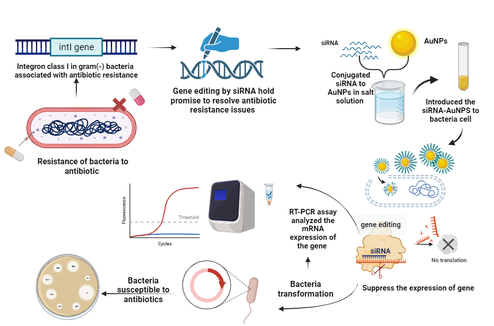
Background: This study explores the application of gold nanoparticles (AuNPs) as a delivery tool for nucleic acids, specifically small interfering RNA (siRNA), in genetic engineering to achieve gene silencing. Methods: This study act on utilizes commercially available AuNPs, characterizing them through techniques such as Field Emission Scanning Electron Microscopy and X-ray diffraction. The study focused on the impact of these AuNPs on delivering siRNA to bacterial cells, particularly Pseudomonas aeruginosa, without the need for additional conjugation compounds. Results: The results show the AuNPs were homogeneously shaped with smaller-sized particles exhibiting a nearly spherical form, and their size increased from 5nm to larger sizes. Various concentrations of AuNPs did not affect the viability of P. aeruginosa. The efficiency of conjugation was confirmed using gel documentation, and the silencing of the antibiotic resistance integron gene (intI) in P. aeruginosa was assessed through a plating method. The results demonstrated a high transformation frequency, with a significant percentage (100%) of colonies showing complete transformation from resistance to sensitivity. Conclusion: The findings indicated the potential of using AuNPs as a delivery system a promising approach for gene silencing in genetic engineering, particularly for combating antibiotic resistance in bacteria.
Disclaimer: This is not the final version of the article. Changes may occur when the manuscript is published in its final format.