
APA Style
Batool Abedrabbo, Noor Alhaddad, Jody Aly, Juan Fernando Castaño, Zhansaya Makhambetova, Ken-ichiro Kamei. (2026). Synthetic Polymer Based Nano Drug Delivery Systems for Precise Alzheimer’s Disease Therapy. Cell Therapy & Engineering Connect, 2 (Article ID: 0010). https://doi.org/10.69709/CTEC.2026.104050MLA Style
Batool Abedrabbo, Noor Alhaddad, Jody Aly, Juan Fernando Castaño, Zhansaya Makhambetova, Ken-ichiro Kamei. "Synthetic Polymer Based Nano Drug Delivery Systems for Precise Alzheimer’s Disease Therapy". Cell Therapy & Engineering Connect, vol. 2, 2026, Article ID: 0010, https://doi.org/10.69709/CTEC.2026.104050.Chicago Style
Batool Abedrabbo, Noor Alhaddad, Jody Aly, Juan Fernando Castaño, Zhansaya Makhambetova, Ken-ichiro Kamei. 2026. "Synthetic Polymer Based Nano Drug Delivery Systems for Precise Alzheimer’s Disease Therapy." Cell Therapy & Engineering Connect 2 (2026): 0010. https://doi.org/10.69709/CTEC.2026.104050.
ACCESS
Review Article
Volume 2, Article ID: 2026.0010

Batool Abedrabbo
ba2255@nyu.edu
Noor Alhaddad
nta4054@nyu.edu
Jody Aly
ja4534@nyu.edu
Juan Fernando Castaño
jfc9577@nyu.edu
Zhansaya Makhambetova
zm2285@nyu.edu
Ken-ichiro Kamei
kk4801@nyu.edu
1 Nanobiotechnology Class, Program of Bioengineering, Division of Engineering, New York University Abu Dhabi, Abu Dhabi P.O. Box 129188, UAE
2 Program of Biology, Division of Science, New York University Abu Dhabi, Abu Dhabi P.O. Box 129188, UAE
3 Department of Biomedical Engineering, Tandon School of Engineering, New York University, Brooklyn, NY 11201, USA
4 Department of Biology, Faculty of Arts & Science, New York University, New York, NY 10003, USA
5 Institute for Integrated Cell-Material Sciences (WPI-iCeMS), Institute for Advanced Study, Kyoto University, Yoshida-Ushinomiya-cho, Sakyo-ku, Kyoto 606-8501, Japan
* Author to whom correspondence should be addressed
† These authors contributed equally.
Received: 27 Sep 2025 Accepted: 20 Apr 2026 Available Online: 21 Apr 2026 Published: 13 May 2026
Alzheimer’s disease (AD) remains a major global health challenge due to its multifactorial pathology and the limited efficacy of current therapies, which mainly provide symptomatic relief rather than modifying disease progression. A central obstacle to effective treatment is the blood–brain barrier (BBB), which severely restricts the delivery of therapeutics to the central nervous system (CNS). In this context, synthetic polymer-based nanoparticles have emerged as versatile platforms capable of enhancing drug stability, prolonging systemic circulation, and enabling controlled and targeted delivery across the BBB. This review summarizes recent advances in polymeric nanocarriers for Alzheimer’s disease (AD) therapy, with an emphasis on systems with translational potential, including poly(lactic-co-glycolic acid) (PLGA), poly(ε-caprolactone) (PCL), poly(lactic acid)–poly(ethylene glycol) (PLA–PEG), and polyethylenimine (PEI). Polymer chemistry, degradation kinetics, and surface functionalization can be designed to overcome key drug delivery barriers, while inherent trade-offs are critically addressed, including the slow biodegradation and clearance limitations associated with PCL, as well as the balance between cytotoxicity and transfection efficiency observed in PEI-based systems. Emerging polymeric architectures, including dendrimers and hybrid systems, are also highlighted for their potential to enable multifunctional and tunable drug delivery strategies. The increasing role of advanced human-relevant in vitro models, such as brain organoids and blood–brain barrier (BBB)-on-a-chip platforms, is emphasized, supported by recent regulatory initiatives promoting New Approach Methodologies (NAMs). These systems provide more predictive tools for evaluating nanoparticle transport, safety, and therapeutic response, thereby improving confidence in clinical translation. Collectively, this review suggests that the successful clinical translation of polymer-based nanotherapies for Alzheimer’s disease will depend on the development of safe, scalable, and well-characterized polymer-based systems validated in advanced human-relevant models, alongside early consideration of manufacturability and regulatory alignment.
Alzheimer’s disease (AD) is a gradual and irreversible neurodegenerative disease, characterized by cognitive, memory, and behavioral impairments that ultimately lead to total dependence on caregivers [1]. Globally, the number of new cases of Alzheimer’s disease and other dementias reached 7.24 million in 2019, representing a 147.95% increase since 1990, making Alzheimer’s disease the most prevalent form of dementia and a major global health concern [2]. In the United Arab Emirates (UAE) and the broader Middle East and North Africa (MENA) region, Alzheimer’s disease (AD) is emerging as a major public health concern. As of 2019, the age-standardized prevalence of dementia in the MENA region was 777.6 per 100,000 population, representing a 3.0% increase compared with 1990 [3]. This rise has been attributed to an aging population, improved diagnostic rates, and the high prevalence of risk factors such as hypertension. Pathologically, AD is characterized by two principal hallmarks: extracellular amyloid-β (Aβ) plaques and intracellular neurofibrillary tangles (NFTs). Aβ plaques result from aberrant cleavage of the amyloid precursor protein (APP) by β-secretase (BACE1) and γ-secretase, producing various Aβ isoforms, notably the aggregation-prone Aβ42 [4,5]. Neurofibrillary tangles (NFTs) arise when tau protein undergoes hyperphosphorylation, leading to microtubule destabilization and subsequent impairment of axonal transport [6]. This tau pathology progresses in a characteristic and predictable sequence, known as Braak staging, beginning in the entorhinal cortex and hippocampus and subsequently spreading to neocortical regions. Alzheimer’s disease is broadly classified into two main types: sporadic Alzheimer’s disease (SAD) and familial Alzheimer’s disease (FAD). Sporadic AD, also referred to as late-onset AD, is the most prevalent form and is influenced by a complex interplay of genetic predisposition and environmental factors [7]. Although no single gene directly causes SAD, the apolipoprotein E (APOE) ε4 allele significantly increases risk by elevating Aβ levels and promoting neuroinflammation [8]. The APOE gene, which is involved in lipid metabolism and amyloid-β (Aβ) clearance, contributes to elevated Aβ accumulation, neuroinflammation, and oxidative stress in carriers of the ε4 allele. Additional genes identified through genome-wide association studies, including triggering receptor expressed on myeloid cells 2 (TREM2), Clusterin (CLU), phosphatidylinositol-binding clathrin assembly protein (PICALM), and bridging integrator 1 (BIN1), also modulate amyloid-β (Aβ) metabolism, tau protein processing, lipid metabolism, and neuroinflammatory pathways [6]. Risk factors, including hypertension, diabetes, obesity, and traumatic brain injury, can contribute to disease progression by exacerbating vascular dysfunction, impairing amyloid-β (Aβ) clearance, and promoting chronic neuroinflammation [7,9]. By contrast, FAD (early-onset AD) accounts for a smaller fraction of AD cases and typically follows an autosomal dominant pattern. Mutations in the APP gene near β- and γ-secretase sites increase the production of toxic Aβ42, while variants in presenilin 1 (PSEN1) and presenilin 2 (PSEN2) disrupt γ-secretase function, further increasing the Aβ42-to-Aβ40 ratio [10]. These mutations accelerate amyloidosis and tauopathy, leading to pronounced neurodegeneration. Current therapeutic approaches for Alzheimer’s disease (AD) primarily include cholinesterase inhibitors (ChEIs) and an N-methyl-D-aspartate (NMDA) receptor antagonist. These treatments provide symptomatic relief but do not alter disease progression. ChEIs, such as donepezil, rivastigmine, and galantamine, act by inhibiting the breakdown of acetylcholine, thereby enhancing cholinergic neurotransmission. In contrast, memantine reduces glutamate-mediated excitotoxicity through NMDA receptor antagonism [11-14]. Despite their clinical utility, these drugs do not address the underlying pathophysiology or prevent progressive neuronal damage, and their adverse effects can hinder long-term adherence [15]. A major challenge in developing more effective therapies is the blood–brain barrier (BBB; Figure 1), a selective physiological interface that limits the delivery of therapeutics to the central nervous system. This restriction significantly hinders the transport of potential disease-modifying agents, including those targeting amyloid-β (Aβ) plaques and tau tangles [16-18]. For example, cholinesterase inhibitors (ChEIs) are commonly associated with gastrointestinal disturbances, while memantine may cause dizziness and confusion [19]. Most importantly, no existing drug effectively modifies the core pathological processes of AD, revealing a critical need for novel strategies. Nanoparticle (NP) technology offers promising solutions by harnessing nanoscale materials to improve the targeting, uptake, and bioavailability of therapeutic compounds within the brain [15, 20-24]. Lipid-based systems, such as liposomes and solid lipid nanoparticles, are biocompatible and simple to fabricate but offer limited long-term drug release, posing challenges in chronic AD treatment. Exosomes exhibit low immunogenicity and possess inherent targeting capabilities; however, challenges related to large-scale isolation and their intrinsic heterogeneity limit their clinical applicability [25,26]. Metallic nanoparticles, such as gold and iron oxide, are highly effective in imaging and theranostic applications; however, their slow biodegradation can lead to tissue accumulation, raising safety concerns associated with prolonged use [27,28]. In contrast, synthetic polymeric nanoparticles composed of biodegradable materials such as poly(lactic-co-glycolic acid) (PLGA), poly(ε-caprolactone) (PCL), and poly(lactic acid)–poly(ethylene glycol) (PLA–PEG) offer significant advantages for Alzheimer’s disease therapy. Their composition can be engineered to modulate drug release profiles, enabling sustained delivery over several weeks, which is particularly important in the context of a progressively degenerative disease. Polymer degradation yields non-toxic byproducts (e.g., lactic and glycolic acids), reducing long-term toxicity. Surface modifications may enable specific binding to Aβ or hyperphosphorylated tau, improving blood–brain barrier penetration and minimizing off-target effects. Polymeric nanoparticles exhibit lower immunogenicity than metallic or lipid-based formulations, thereby further enhancing their safety profile. As a result, they represent promising delivery vehicles for a wide range of therapeutic agents, including biologics and gene-editing tools, in the treatment of Alzheimer’s disease (AD). Given the limitations of current AD therapies and the challenges posed by the BBB, synthetic polymeric nanoparticles present a promising strategy for targeted, sustained drug delivery. Their tunable release profiles, biocompatibility, and potential for surface functionalization enable enhanced penetration into brain tissue, making them particularly well-suited to addressing the multifaceted pathology of AD. Accordingly, this review focuses on the design, physicochemical properties, and therapeutic potential of synthetic polymeric nanoparticles, with particular emphasis on their ability to overcome existing therapeutic barriers. It further examines how these systems may facilitate the development of more effective, disease-modifying strategies for Alzheimer’s disease (AD).
This review was conducted as a narrative literature review rather than a systematic review. Relevant publications were identified through comprehensive searches of PubMed, Google Scholar, Scopus, and Google Search, supplemented by manual screening of the reference lists of key articles to identify additional relevant studies not retrieved during the initial searches. Searches were conducted using combinations of keywords including, but not limited to, Alzheimer’s disease, polymeric nanoparticles, synthetic polymers, drug delivery, gene delivery, blood–brain barrier, PLGA, PCL, PLA–PEG, polyethylenimine, dendrimers, brain organoids, and blood–brain barrier (BBB)-on-a-chip. Only publications written in English were included. In total, approximately 200 articles were retrieved and reviewed in full text. Study selection was guided primarily by relevance to the topic, scientific rigor, and recency, with particular emphasis on publications published between 2018 and 2025. No predefined inclusion or exclusion criteria were applied, consistent with the narrative design of this review; however, articles that were peripheral to the scope of polymer-based nanocarriers, lacked sufficient methodological information, or were redundant with more recent or comprehensive publications were excluded during the screening process. Accordingly, only a subset of the initially screened articles was included and cited in the final manuscript. Among the cited references, approximately 43% were original experimental research articles, whereas 57% were review articles, reflecting a balance between primary mechanistic studies and integrative perspectives. All cited works were available as full-text articles and were reviewed in their entirety. A small number of included publications were authored or co-authored by the present review’s authors; however, these were included only when directly relevant to the topic and were assessed using the same selection and evaluation criteria applied to all other sources.
Nanomedicine began to emerge in the mid-20th century with the exploration of nanoparticles (NPs) for biomedical applications. A key milestone was achieved in the 1960s, when Alec Bangham and colleagues first described liposomes—phospholipid vesicles that subsequently formed the basis for nanoparticle-based drug delivery systems [29]. In 1986, Matsumura et al. formally characterized the enhanced permeability and retention (EPR) effect, describing how the unique pathophysiological features of solid tumor vasculature enable the preferential accumulation of NPs [30]. By the late 1980s, the introduction of poly(ethylene glycol) (PEG) coatings on NPs significantly improved their ability to evade immune surveillance, thereby extending circulation time [31,32]. A major milestone in nanomedicine was reached in 1995, when the U.S. Food and Drug Administration (FDA) approved Doxil, a PEGylated liposomal formulation of doxorubicin, for the treatment of Kaposi’s sarcoma [33]. This approval marked a significant breakthrough in NP-based drug delivery and demonstrated the clinical feasibility of nanoscale therapeutic. The clinical success of Doxil highlighted the potential of nanoparticle-based drug delivery systems, thereby stimulating efforts to extend similar strategies for the treatment of neurodegenerative diseases. As Alzheimer’s disease (AD) became increasingly recognized as a major neurodegenerative disorder, research efforts progressively shifted toward addressing the challenge of delivering therapeutics across the blood–brain barrier (BBB). Advances in molecular biology identified specific targets such as Aβ plaques, highlighting the need for precise drug delivery into the brain [34]. By the early 2000s, efforts had intensified toward designing nanoparticles capable of crossing the blood–brain barrier (BBB), leveraging receptor-mediated transcytosis and targeting regions where barrier integrity may be compromised in Alzheimer’s disease pathology. During this period, researchers began functionalizing nanoparticles with ligands that selectively bind Aβ plaques, enabling targeted drug delivery [35,36]. Polymer-based nanoparticles, particularly those composed of biodegradable materials such as poly(lactic-co-glycolic acid) (PLGA) [37] and poly(ε-caprolactone) (PCL) [38], gained considerable attention due to their physicochemical stability and capacity for controlled and sustained drug release. These properties are particularly advantageous in AD models, where prolonged drug retention within the brain may improve therapeutic efficacy and contribute to slowing disease progression. The versatility of polymeric nanoparticles further enhances their appeal, as they can encapsulate a wide range of therapeutic agents, including small molecules, proteins, and nucleic acids [29]. For example, curcumin-loaded PLGA nanoparticles have shown promise in reducing amyloid aggregation and oxidative stress [35]. However, challenges related to bioavailability persist, and ongoing research is investigating nanomaterial-based delivery systems—such as lipid nanoparticles and micelles—to improve curcumin’s solubility, stability, and ability to cross the blood–brain barrier (BBB), while minimizing toxicity.
The blood–brain barrier (BBB) is a highly selective, semipermeable interface formed by endothelial cells that prevents circulating solutes from non-selectively entering the extracellular fluid of the central nervous system (CNS), where neurons are located (Figure 1) [16-18]. The BBB is formed by endothelial cells of the capillary wall, astrocyte end-feet ensheathing the capillary, and pericytes embedded in the capillary basement membrane [39]. The tight junctions between endothelial cells constitute a critical structural component of the blood–brain barrier (BBB), significantly restricting the passage of substances from the bloodstream into the brain. Although this barrier is essential for protecting the central nervous system (CNS) from toxins and pathogens, it simultaneously presents a major obstacle to the delivery of therapeutic agents to the brain. For a drug to cross the BBB, it generally needs to be of low molecular weight(typically under 400–600 Da) and lipid soluble to diffuse across the cell membranes [16,40]. However, nanoparticles can circumvent this barrier through several mechanisms. Passive diffusion enables the transport of small, lipophilic molecules across the blood–brain barrier, whereas receptor-mediated transcytosis facilitates transport through ligand–receptor interactions. In particular, targeting ligands such as transferrin or insulin can be employed to engage specific receptors on the BBB and promote transcellular delivery [41-45]. Adsorptive-mediated transcytosis exploits electrostatic interactions between positively charged nanoparticles and the negatively charged BBB surface, facilitating uptake and transport [46]. Among nanoparticle-based strategies, polymeric nanoparticles are particularly effective owing to their biocompatibility, tunable physicochemical properties, and capacity to encapsulate a wide range of therapeutic agents. Functionalization may enable targeted drug delivery, particularly for therapeutic strategies targeting Aβ plaques and tau tangles [47]. One notable strategy is Angiopep-2–mediated transport, in which Angiopep-2 binds to low-density lipoprotein receptor-related protein 1 (LRP1) expressed on endothelial cells. This interaction triggers receptor-mediated endocytosis and subsequent intracellular transport, ultimately facilitating the release of the therapeutic payload into brain tissue (Figure 2) [48]. Given that low-density lipoprotein receptor-related protein 1 (LRP1) is involved in amyloid-β (Aβ) clearance, Angiopep-2–modified nanoparticles can enhance targeted drug delivery to affected brain regions. This strategy may improve therapeutic uptake across the blood–brain barrier and potentially reduce both Aβ aggregation and tau pathology. Nose-to-brain drug delivery provides a non-invasive alternative route that bypasses the blood–brain barrier (BBB) by using the nasal cavity as a direct route for the transport of therapeutic agents to the central nervous system (CNS) [49,50]. This pathway may minimize systemic absorption while supporting targeted delivery. Nanoparticles can reach the brain via the olfactory route, in which they penetrate the olfactory epithelium and are transported along the olfactory nerve to the olfactory bulb, or via the trigeminal nerve pathway, where they are absorbed through the respiratory epithelium and subsequently transported to deeper brain regions. To enhance drug retention and absorption, nanoparticles (NPs) can be engineered for mucoadhesion, thereby reducing rapid mucociliary clearance and enabling controlled release for sustained therapeutic effects [51]. Transport mechanisms, such as paracellular diffusion and transcellular transport, further facilitate their passage between epithelial cells [52,53]. Together, these strategies may make nanoparticle systems effective for delivering anti-inflammatory agents aimed at mitigating AD-related neuroinflammation [53]. Despite its potential, nose-to-brain delivery faces several challenges, including mucociliary clearance, which reduces drug residence time in the nasal cavity, and the formation of protein coronas, which can alter nanoparticle behavior and promote off-target interactions [54-56]. Nevertheless, polymeric nanoparticles, such as poly(lactic-co-glycolic acid) (PLGA) and chitosan-based systems, along with lipid nanoparticles, nanoemulsions, and dendrimers, have demonstrated considerable promise in protecting therapeutic agents from enzymatic degradation while enhancing absorption and improving overall bioavailability [20]. Both BBB delivery and nose-to-brain delivery offer promising strategies for nanoparticle-based AD therapies. While BBB-targeted approaches enable precise drug transport, nose-to-brain delivery provides a non-invasive route to the CNS. Continued advances in nanoparticle engineering and optimization of delivery mechanisms will be essential for enhancing therapeutic efficacy in the treatment of Alzheimer’s disease (AD).
Several synthetic polymeric nanocarriers have been developed to enhance drug delivery in Alzheimer’s disease (AD). These systems leverage biocompatible, biodegradable polymers to encapsulate therapeutic agents, protect them from degradation, and release them in a controlled manner. Crucially, many polymeric nanoparticles (NPs) can be engineered to cross the blood–brain barrier (BBB) through size optimization, surface modification (e.g., PEGylation or attachment of targeting ligands), and tuning of polymer properties such as hydrophobicity and degradation rate [57]. In general, polymeric NPs offer sustained drug release, improved bioavailability in the brain, and reduced systemic toxicity compared with free drugs [58,59]. Below, key classes of synthetic polymeric nanoparticles (NPs) used in Alzheimer’s disease (AD) therapy are discussed, including PLGA, PCL, PLA–PEG copolymers, PEI-based polyplexes, and emerging polymeric systems, with emphasis on recent advances (2023–2025) in their design, therapeutic mechanisms, and clinical relevance (Figure 3). 5.1. Poly(lactic-co-glycolic acid) (PLGA) Poly(lactic-co-glycolic acid) (PLGA) is an FDA-approved, linear copolymer widely used in nanomedicine. Its application in neuro-nanomedicine is primarily attributed to its excellent biocompatibility, predictable hydrolytic degradation, and versatility in formulation with both hydrophilic and hydrophobic therapeutic payloads (Figure 3) [60-67]. By adjusting the lactic-to-glycolic acid ratio, researchers can precisely tune key physicochemical properties such as glass-transition temperature, crystallinity, and, consequently, drug release kinetics. For example, a 50:50 ratio typically results in degradation within approximately one week, whereas poly(lactic acid) (PLA)-rich formulations may persist in vivo for more than 18 weeks [64,68]. Surface modification (e.g., PEGylation, ligand conjugation) further extends circulation time and enables receptor-mediated BBB transport [64]. Its low toxicity profile, combined with its excellent compatibility with other polymers and functional additives [69], makes it an ideal candidate for drug encapsulation applications [63-64, 68]. Notably, recent studies indicate that PLGA itself may exert therapeutic effects beyond serving as a passive carrier [62,70]. Paul et al. demonstrated that drug-free (“native”) poly(lactic-co-glycolic acid) (PLGA) nanoparticles (NPs) can inhibit amyloid-β (Aβ42) fibrillization and provide neuroprotective effects on cortical neurons. This unexpected intrinsic activity suggests PLGA NPs might help ameliorate AD pathology by “soaking up” or interfering with toxic Aβ oligomers and tau aggregates [62]. Additionally, PLGA NPs are being investigated for gene delivery in AD. For instance, cationic-surface PLGA NPs loaded with siRNA against Cdkn2a (p16INK4a) have been shown to successfully silence this aging-associated gene in microglia. This gene modulation effectively “rejuvenated” microglial cells, enhanced their amyloid-β (Aβ) phagocytic activity, and reduced amyloid plaque burden in AD mouse models [71]. This strategy was associated with improved cognitive outcomes, highlighting how PLGA nanocarriers can be tailored for immunomodulation in AD [72]. Overall, PLGA-based nanoparticles represent a versatile platform that combines a favorable safety profile—owing to their degradation into endogenous metabolites such as lactic acid and glycolic acid—with the flexibility to encapsulate a wide range of therapeutic payloads. Notably, emerging evidence also suggests that PLGA nanoparticles may themselves exert beneficial biological effects, including potential disease-modifying properties. Tracy et al. demonstrated that 200 nm poly(lactic-co-glycolic acid)-block-hyaluronic acid (PLGA-b-HA) nanoparticles preferentially accumulate in the hippocampus of 13–16-month-old APP/PS1 mice, as well as in age-matched wild-type mice. This enhanced accumulation was attributed to increased blood–brain barrier (BBB) permeability associated with aging. The HA corona binds the CD44 receptor on reactive astrocytes and activated microglia, providing dual benefits of (i) passive entry through “leaky” vasculature and (ii) active cellular uptake [73]. 5.2. Poly(ε-caprolactone) (PCL) Polycaprolactone (PCL) is another FDA-approved biodegradable polyester used in nanoparticle formulation [74], distinguished by its more hydrophobic nature and slower degradation kinetics compared with that of PLGA (Figure 3) [75]. These properties make PCL particularly attractive for applications requiring long-term, sustained drug release, although they also raise important questions regarding long-term polymer residence and biocompatibility in the central nervous system. Early work by Mahmoudi et al. exploited the slow degradation profile of PCL by encapsulating memantine (an NMDA receptor antagonist) within PCL nanocapsules (MEM@PCL), which improved pharmacokinetics and reduced off-target toxicity [76]. Subsequently, the same group developed 7-methoxytacrine-loaded PCL nanocapsules, demonstrating controlled release of this rapidly metabolized cholinesterase inhibitor and improved in vitro neuroprotection [77]. More recently, Müller et al. developed hot-melt-extruded poly(ε-caprolactone) (PCL) matrices incorporating 1–50 wt% galantamine hydrobromide, achieving near zero-order release kinetics. This sustained-release profile is particularly advantageous for ultra-long-acting oral therapeutic applications [78]. Beyond payload formulation, surface engineering has emerged as a powerful strategy to overcome PCL’s intrinsic limitations in BBB transport. Gu et al. adopted a biomimetic approach, cloaking PCL nanoparticles with erythrocyte membranes and grafting the BBB-targeting peptide TGNYKALHPHN (TGN) [79]. These TGN-RBC-NPs significantly enhanced brain accumulation of curcumin, reduced neuroinflammation, preserved hippocampal neurons, and improved cognitive performance in Alzheimer’s disease mouse models, thereby underscoring the critical role of surface functionalization in improving central nervous system (CNS) drug delivery. Despite these advances, PCL nanoparticles persist longer in biological systems than faster-degrading polymers, and their long-term fate in the brain, particularly under chronic dosing, is not yet fully established. Current in vivo tracking studies are beginning to elucidate PCL nanoparticle biodegradation and clearance pathways; however, definitive evidence on potential accumulation of residual polymer or metabolites in neural tissue is still lacking. Accordingly, establishing the long-term safety profile of poly(ε-caprolactone) (PCL) nanocarriers under chronic use conditions represents a critical prerequisite for their broader clinical translation. To address concerns related to the slow degradation and prolonged tissue residence of poly(ε-caprolactone) (PCL), several material engineering strategies have been explored. One common approach is copolymerization, such as blending PCL with faster-degrading polyesters, including poly(lactic acid) (PLA) [80-82] or poly(lactic-co-glycolic acid) (PLGA) [83]. This strategy accelerates hydrolytic degradation while maintaining the mechanical stability and sustained-release properties of PCL [84]. The incorporation of hydrophilic blocks (e.g., PEG) or ester-rich segments increases water uptake and enhances enzymatic accessibility, thereby shortening in vivo residence time. Additional strategies include reducing molecular weight [85], introducing amorphous domains [86], and formulating PCL-based nanocomposites with biodegradable additives [86]. These approaches collectively modulate degradation kinetics without compromising drug-loading capacity. These strategies collectively seek to balance the inherent advantage of poly(ε-caprolactone) (PCL) in enabling ultra-sustained drug release with improved long-term biocompatibility, a key consideration for chronic neurodegenerative conditions such as Alzheimer’s disease. Collectively, these advances position PCL nanoparticles as a valuable complement to PLGA-based systems, offering ultra-sustained release and versatile surface modification capabilities for central nervous system therapeutics, provided that future studies confirm their long-term clearance and safety. 5.3. Poly(lactic acid)-poly(ethylene glycol) (PLA-PEG) PLA-PEG is a block copolymer comprising a hydrophilic polyethylene glycol (PEG) segment and a hydrophobic poly(lactic acid) (PLA) segment. This amphiphilic architecture provides a dual advantage by enabling prolonged systemic circulation while also facilitating efficient drug encapsulation (Figure 3) [87]. Typical PLA–PEG nanoparticles fall within the 50–200 nm range, which is sufficiently small to evade the reticuloendothelial system while remaining large enough to avoid rapid renal clearance, and within the optimal size window for transcytosis across the blood–brain barrier (BBB) [88]. The PEG “stealth” layer reduces opsonization and hepatic clearance, while the PLA block enables the encapsulation of hydrophobic drugs within a biodegradable matrix. To facilitate transport across the blood–brain barrier (BBB), researchers have functionalized the PEG shell with targeting ligands such as Angiopep-2 or transferrin, thereby exploiting receptor-mediated transcytosis mechanisms. Such surface modifications have been reported to increase brain uptake by approximately three- to ten-fold [42, 61, 89]. Therapeutically, ligand-decorated PLA-PEG carriers have excelled in Alzheimer’s disease (AD) models. Donepezil-loaded PLGA-b-PEG NPs prolonged drug exposure, suppressed acetylcholinesterase more effectively than free donepezil, and improved Morris-water-maze performance in Aβ-infused rats [90]. Flash-nanoprecipitated ibuprofen-loaded PLA–PEG nanoparticles demonstrated stability for up to 34 days and increased cerebral ibuprofen concentrations by approximately fourfold, thereby attenuating microglial activation in vivo [79]. Beyond pharmacology, combining NGF-loaded PEG-PLGA NPs with neural-stem-cell transplantation restored cholinergic neurons and rescued cognition in AD rats, underscoring the platform’s compatibility with regenerative approaches [91]. 5.4. Polyethylenimine (PEI) Polyethylenimine (PEI) is a highly cationic polymer characterized by a high density of primary, secondary, and tertiary amine groups, which impart strong proton-buffering capacity and robust affinity for nucleic acid binding (Figure 3) [92-95]. These properties underpin PEI’s long-standing utility as one of the most efficient non-viral vectors for gene delivery, particularly for siRNA and plasmid DNA. Both linear and branched polyethylenimine chains can be engineered into dendrimeric or globular architectures, resulting in nanoparticles with well-defined sizes (typically 20–200 nm), high surface-area-to-volume ratios, and multivalent amine functionality. These physicochemical properties are advantageous for intracellular delivery applications, particularly in neurodegenerative disease models, including Alzheimer’s disease (AD) [96]. Functionalization of PEI-based nanocarriers with targeting ligands—such as apolipoprotein E (ApoE), transferrin, or blood–brain barrier (BBB) shuttle peptides—facilitates receptor-mediated transcytosis, thereby enhancing their transport across the BBB and improving brain uptake efficiency [53]. Following cellular internalization, PEI’s strong cationic charge facilitates endosomal escape through the so-called “proton-sponge” effect, a critical requirement for efficient cytosolic delivery of nucleic acid therapeutics [97,98]. Consistent with this mechanism, Zhang et al. reported approximately 88% knockdown of Bace1 following intraventricular administration of a PEI-based siRNA complex. This intervention resulted in an approximately 60% reduction in soluble amyloid-β (Aβ) levels and produced significant cognitive improvement in 5×FAD mouse models [99]. Despite these advantages, cytotoxicity remains the primary limitation of high-molecular-weight or highly branched PEI, largely due to excessive membrane disruption and nonspecific electrostatic interactions with cellular components [100]. Consequently, substantial efforts have focused on mitigating PEI-associated toxicity while preserving its high transfection efficiency, leading to a well-recognized trade-off between biocompatibility and delivery performance [101]. PEGylation is one of the most widely adopted strategies to attenuate PEI cytotoxicity by partially shielding surface charges and reducing nonspecific protein adsorption [102]. Experimental studies have demonstrated that moderate PEG grafting significantly enhances cell viability and serum stability. However, excessive PEGylation can impair cellular uptake and endosomal escape, thereby reducing transfection efficiency [103]. These findings underscore the importance of carefully tuning PEG chain length and grafting density to balance safety and gene delivery efficacy. Charge-attenuation strategies, including partial acetylation of PEI amine groups, have been explored as an approach to reduce cytotoxicity. Controlled acetylation can mitigate membrane disruption and, in some cases, modulate polymer–nucleic acid interactions to promote more efficient intracellular payload release. As a result, transfection efficiency may be maintained or even enhanced at intermediate degrees of modification [104]. In contrast, excessive neutralization can compromise nucleic acid condensation and cellular uptake, again highlighting the existence of an optimal modification window [105]. More recently, zwitterionic shielding strategies [106], such as the grafting of sulfobetaine or carboxybetaine moieties onto PEI, have emerged as effective approaches for suppressing protein fouling and cytotoxicity while preserving nucleic acid binding capability [107]. Original experimental studies have demonstrated that mildly zwitterion-modified PEI complexes exhibit substantially reduced cytotoxicity while retaining effective gene-silencing activity. In contrast, excessive surface shielding can impair cellular uptake and consequently reduce transfection efficiency. An orthogonal and complementary approach involves the use of low-molecular-weight (LMW) PEI, which inherently exhibits reduced cytotoxicity but also lower transfection efficiency [108]. To address this limitation, low-molecular-weight (LMW) PEI units have been engineered into biodegradable or reversibly crosslinked network structures. These systems retain the nucleic acid condensation efficiency characteristic of high-molecular-weight PEI while undergoing intracellular degradation into less toxic fragments, thereby improving overall biocompatibility. Such systems have demonstrated markedly improved safety profiles with only moderate reductions in transfection efficiency, substantially improving the overall therapeutic index [94,95]. Collectively, these modification strategies establish PEI as a highly tunable gene delivery platform, in which cytotoxicity and transfection efficiency can be rationally balanced through molecular engineering. Rather than being viewed only as an inherent limitation, the toxicity–efficacy trade-off associated with PEI has become an important design consideration. This perspective has enabled the rational development of safer, application-specific PEI-based nanocarriers for nucleic acid delivery in neurodegenerative diseases, as well as in broader biomedical applications.
Although the four polymers discussed—PLGA, PCL, PLA–PEG, and PEI—share the general function of drug and gene delivery, a more detailed analysis reveals important differences in their mechanisms of action, optimal applications, and translational potential. The selection of a polymer is not arbitrary; rather, it represents a strategic decision that significantly influences the therapeutic outcome [109]. 6.1. Mechanistic Differences in Drug Encapsulation and Release Kinetics The ability to load and release a therapeutic agent is one of the most important functions of a nanocarrier. Here, the physicochemical properties of the polymers dictate their performance (Table 1). Comparative analysis of polymeric nanocarriers for AD therapy. PLGA [110] and PCL [111], both hydrophobic polyesters, are particularly effective for encapsulating hydrophobic drugs using methods such as nanoprecipitation or emulsion evaporation. The drug is typically entrapped within the solid polymer matrix. The release is then governed by a combination of drug diffusion and polymer degradation. PLGA, with its amorphous structure and tunable lactic-to-glycolic acid ratio, generally exhibits a characteristic biphasic release: an initial “burst release” of surface-adsorbed drug, followed by a slower, sustained release as the polymer matrix erodes [112,113]. This can be beneficial for therapies requiring an initial high dose followed by a maintenance dose. Because of its semi-crystalline nature, PCL and slower degradation, offers a more linear, zero-order release profile over much longer periods (months to years) [114], making it ideal for neuroprotective agents that require a constant, steady-state concentration in the brain. PLA-PEG, as a diblock copolymer, forms core–shell nanoparticles in which the hydrophobic poly(lactic acid) (PLA) core serves as the principal compartment for drug encapsulation, analogous to poly(lactic-co-glycolic acid) (PLGA) and poly(ε-caprolactone) (PCL)-based systems. However, the hydrophilic PEG shell can be used to conjugate or adsorb hydrophilic drugs or biologics, creating a dual-loading capacity [115]. The release profile is generally comparable to that of PLGA-based systems; however, it can be further fine-tuned by adjusting the length of the polyethylene glycol (PEG) chain. This parameter influences nanoparticle hydrophilicity and its interactions with the surrounding aqueous environment, thereby modulating drug release kinetics. PEI operates through a fundamentally different mechanism, as it is primarily engineered for gene delivery rather than for the controlled delivery of small-molecule drugs. Its highly cationic nature allows PEI to strongly bind, compact, and condense negatively charged nucleic acids (such as siRNA or plasmid DNA), forming polyplexes through electrostatic interactions [92-93, 116]. Within the intracellular environment, release of the genetic payload is not governed by gradual diffusion but occurs as an “on-demand” event, primarily triggered by the proton sponge effect within endosomes [97,98]. The buffering capacity of protonatable amines in PEI leads to the accumulation of protons and counter-ions, resulting in osmotic swelling and subsequent endosomal rupture, thereby facilitating the release of nucleic acid cargo into the cytosol [117]. This mechanism is highly effective for gene delivery; however, it is poorly suited for the sustained release of conventional small-molecule drugs, for which a slow and predictable elution profile over time is typically required. 6.2. Navigating the Blood–Brain Barrier: Stealth, Charge, and Targeting Crossing the blood–brain barrier (BBB) represents the principal rate-limiting step for most central nervous system (CNS) therapeutics. Accordingly, different polymer-based delivery systems employ distinct strategies to facilitate BBB penetration and enhance brain targeting efficiency. PLA-PEG is widely recognized as an effective “stealth” delivery system. The dense layer of hydrophilic PEG on the nanoparticle surface effectively shields it from opsonization and clearance by the reticuloendothelial system. This significantly increases its circulation half-life, thereby extending the time window for nanoparticles to interact with and traverse the blood–brain barrier (BBB) [118]. Although the precise mechanism remains under investigation, it is thought to involve a combination of passive diffusion and receptor-mediated transcytosis (RMT), potentially mediated through interactions with receptors such as LRP1 [119]. PLGA and PCL nanoparticles, in their unmodified state, have a more limited ability to cross the BBB [29]. Their passage is largely dependent on their small size and hydrophobic surface properties, which permit a degree of passive diffusion. However, achieving clinically relevant brain concentrations typically necessitates surface functionalization with targeting ligands (e.g., antibodies against the transferrin receptor or peptides such as ApoE) to enable active receptor-mediated transcytosis (RMT). While this approach enhances versatility, it also introduces additional complexity in design and manufacturing [120]. PEI exploits its positive charge as a key functional feature. Its cationic surface interacts with negatively charged components of BBB endothelial cell membranes, thereby facilitating adsorptive-mediated transcytosis [121]. While effective, this mechanism is less specific than RMT and may lead to off-target effects and potential toxicity [116,122]. 6.3. Biocompatibility and Toxicity: The Degradation Dilemma The long-term safety of nanocarriers is a critical consideration, particularly in the context of chronic diseases such as Alzheimer’s disease (AD). PLGA and PCL are generally regarded as highly biocompatible, as they degrade into natural metabolites, including lactic acid, glycolic acid, and 6-hydroxycaproic acid, which are subsequently cleared from the body. However, important considerations remain regarding their in vivo behavior. The degradation of PLGA creates an acidic microenvironment [123], which can be detrimental to the stability of encapsulated drugs and may induce a localized inflammatory response [124]. PCL’s very slow degradation rate, while beneficial for drug release, means that the polymer may persist in the brain for prolonged periods, and the long-term consequences remain unclear. PLA–PEG retains the biocompatibility of PLA, and the PEG component is likewise considered safe and has been incorporated into numerous approved formulations. However, emerging evidence indicates potential concerns regarding the development of anti-PEG antibodies [125,126], which may result in accelerated clearance of nanoparticles upon repeated administration, a phenomenon referred to as accelerated blood clearance (ABC). Among the four polymers discussed, PEI presents the most significant safety concerns. Its high positive charge density, while essential for gene delivery function, is also responsible for its inherent cytotoxicity. PEI can disrupt cellular membranes and induce apoptosis. Although this toxicity can be reduced through the use of low-molecular-weight PEI or via conjugation with other polymers, it remains a major limitation to its clinical translation. In conclusion, the choice of a synthetic polymer for AD therapy is a complex multi-parameter optimization problem. There is no single “best” polymer; rather, an optimal polymer may be selected for a specific therapeutic strategy. Future progress is likely to depend in the development of hybrid systems that integrate the most advantageous properties of each material, thereby enabling the design of more sophisticated and effective “smart” nanotherapies for Alzheimer’s disease.Feature
PLGA
PCL
PLA/PLA-PEG
PEI
Primary Application
Versatile delivery of small molecules & biologics.
Long-term, sustained release of neuroprotective agents.
“Stealth” delivery of sensitive cargo (e.g., antibodies, siRNA).
Gene therapy (siRNA, plasmid DNA delivery).
Drug Compatibility
Primarily hydrophobic drugs within the core matrix.
Primarily hydrophobic drugs; high permeability.
Hydrophobic core + potential for hydrophilic drug conjugation to PEG shell.
Nucleic acids (negative charge) via electrostatic condensation.
Release Mechanism
Biphasic: Initial burst followed by sustained release via bulk erosion.
Zero-order, linear release over extended periods via surface erosion.
Core-shell diffusion and polymer erosion; tunable via PEG length.
Endosomal escape (“proton sponge” effect); triggered release of cargo.
BBB Transport Strategy
Limited passive diffusion; requires active targeting ligands (e.g., ApoE) for efficiency.
Very limited passive diffusion; requires active targeting ligands.
“Stealth” effect from PEG layer prolongs circulation, enhancing the probability of crossing.
Adsorptive-mediated transcytosis due to positive surface charge.
Key Advantage
FDA-approved; highly tunable degradation & release profile.
Excellent for chronic, long-term therapy due to slow, steady release.
Prolonged blood circulation and enhanced BBB penetration.
Highest non-viral gene transfection efficiency.
Primary Challenge
Acidic degradation byproducts can harm labile drugs and cause inflammation.
Very slow degradation (residence time in brain); hydrophobicity limits drug types.
“PEG dilemma” (can hinder cellular uptake); potential for anti-PEG antibodies.
Inherent cytotoxicity due to high positive charge density.
Clinical Development Status
FDA-approved for multiple parenteral drug delivery applications; widely used in approved formulations and clinical trials [64-65, 67].
FDA-approved polymer for biomedical applications; nanoparticle-based drug delivery systems primarily at preclinical and early translational stages [74,86].
FDA-approved (PLA) and clinically validated in multiple drug delivery systems; PLA–PEG block copolymers extensively used in clinical and preclinical nanomedicine [66-67, 87].
Not FDA-approved for systemic drug delivery; extensively investigated in preclinical gene and nucleic acid delivery, with modified forms under translational evaluation [92-95].
Beyond these cornerstone polymers, the field of nanomedicine continues to evolve, through the introduction of novel platforms that offer distinct advantages for central nervous system (CNS) drug delivery. These emerging systems leverage distinct architectures and biological interaction profiles to overcome the challenges of treating Alzheimer’s disease. 7.1. Polyamidoamine (PAMAM) Dendrimers Dendrimers represent a promising class of synthetic polymers with a highly branched, tree-like architecture. Unlike linear polymers, poly(amidoamine) (PAMAM) dendrimers [127,128] are synthesized in a stepwise, layer-by-layer manner, resulting in a highly uniform and spherical nanostructure. This well-defined, monodisperse architecture provides exceptional control over their physicochemical properties. The multivalent surface of PAMAM dendrimers can be densely functionalized with targeting moieties, imaging agents, and therapeutic drugs, creating a highly sophisticated, multifunctional nanodevice. Their unique structure and customizable surface properties have made them effective tools for crossing the blood–brain barrier (BBB), with demonstrated potential for both delivering anti-amyloid agents and exerting intrinsic therapeutic effects by inhibiting protein aggregation. 7.2. Layer-by-Layer Eroding Polyanhydrides Polyanhydrides are a class of biodegradable polymers distinguished by their surface-eroding degradation mechanism. Whereas polymers such as PLGA degrade via bulk hydrolysis, which can lead to an unpredictable burst release, polyanhydrides erode cleanly from the surface inward. This property results in a highly predictable, linear, zero-order drug release, which is ideal for therapies that require a constant, steady-state therapeutic concentration. The FDA-approved use of a polyanhydride wafer implant (Gliadel®) for the treatment of brain tumors has already established a clinical precedent for their safety and utility in the central nervous system (CNS), supporting their potential as promising candidates for future Alzheimer’s disease (AD) therapies [129,130].
A significant challenge in the development of nanotherapies for Alzheimer’s disease lies in the reliance on conventional preclinical models that often have limited ability to predict human responses. Standard two-dimensional cell cultures lack the complex 3D architecture and cellular heterogeneity of the brain, whereas animal models, despite their utility, are constrained by species-specific differences that limit their translational relevance. In response to these limitations, advanced human-relevant in vitro platforms have gained increasing attention, supported by recent initiatives by the U.S. Food and Drug Administration and the National Institutes of Health promoting New Approach Methodologies (NAMs) [131,132]. Brain organoids and organ-on-a-chip systems, including blood–brain barrier models, offer more physiologically relevant environments for evaluating nanoparticle transport, toxicity, and therapeutic efficacy, and are increasingly viewed as complementary tools for translational research and regulatory decision-making. Together, these platforms provide a promising framework for bridging the gap between preclinical development and clinical translation of polymer-based nanomedicines for AD. 8.1. Brain Organoids: Modeling AD Pathology in a Dish Brain organoids are self-assembling, three-dimensional (3D) cultures derived from human pluripotent stem cells (hPSCs) [133-135]. These organoids can recapitulate key aspects of early human brain development, including the formation of distinct brain regions and the presence of diverse cell types such as neurons, astrocytes, and microglia. For AD research, patient-derived hPSCs can be used to generate organoids that intrinsically develop AD-like pathology, such as amyloid-beta aggregation and hyperphosphorylated tau [136,137]. This provides an invaluable human-specific model, enabling researchers to apply polymeric nanoparticles and directly assess their efficacy in reducing amyloid-β (Aβ) plaques or tau tangles within a complex, multicellular environment. Furthermore, the complex cell–cell interactions within these organoids enable a more accurate assessment of nanoparticle-induced neurotoxicity compared with traditional two-dimensional (2D) culture systems [133,138]. 8.2. Organ-on-a-Chip Systems: Recreating the Blood–Brain Barrier The organ-on-a-chip concept refers to a microfluidic device that incorporates living cells within continuously perfused microchannels, thereby recapitulating the physiological and mechanical microenvironment of a human organ at the microscale [139,140]. Of particular relevance to nanomedicine is the BBB-on-a-chip platform, which co-cultures human brain endothelial cells, pericytes, and astrocytes under dynamic flow conditions that replicate blood flow and shear stress. This configuration establishes a functional barrier that more closely resembles the in vivo human blood–brain barrier (BBB) than conventional static Transwell systems, making it particularly valuable for translational research studies [141-143]. These models play a crucial role in the development of nanomedicines. They provide a robust and reproducible means of assessing and quantifying the ability of polymeric nanoparticles to cross the human blood–brain barrier (BBB), with some platforms incorporating microelectrode arrays to measure trans endothelial electrical resistance (TEER) and thereby monitor barrier integrity in real time [144]. Moreover, the live imaging and integrated sensing capabilities of blood–brain barrier-on-a-chip systems enable direct observation of nanoparticle transport pathways, such as receptor-mediated transcytosis versus paracellular leakage, thereby providing mechanistic insights that are critical for optimizing nanocarrier design [143]. The integration of blood–brain barrier-on-a-chip platforms into the nanoparticle development pipeline has the potential to reduce translational risk by generating highly human-relevant preclinical data. When integrated with brain organoid models, these platforms enable more predictive and physiologically relevant assessment of nanoparticle safety, transport efficiency, and therapeutic efficacy, thereby facilitating the accelerated translation of promising nanotherapeutic strategies from preclinical research to clinical application [139,140].
Global regulatory frameworks have progressively adapted to the specific challenges posed by polymer-based nanomedicines without fundamentally altering existing approval pathways. Major agencies, including the FDA, EMA, and PMDA, generally evaluate nano-drug delivery systems under established quality, safety, and efficacy standards, often using combination-product frameworks, while issuing supplementary guidance tailored to nanoscale materials [145]. Early engagement with regulatory authorities is widely encouraged, and reflection papers or product-class–specific guidelines have been developed to support the characterization, manufacturing, and risk assessment of nanomedicines [146-149]. Nevertheless, highly complex or multifunctional polymeric systems may still require case-by-case regulatory evaluation, underscoring the need for continued refinement of regulatory science. In the Middle East and North Africa (MENA) region, regulatory capacity for nanomedicine is heterogeneous but is undergoing rapid development. Historically, fragmented national regulatory approval processes have posed challenges for the introduction of innovative therapies. However, increasing reliance on U.S. Food and Drug Administration (FDA) and European Medicines Agency (EMA) approvals, together with emerging regional harmonization initiatives, is helping to improve access pathways [147]. These developments suggest growing opportunities for advanced polymer-based nano drug delivery systems in MENA, provided that regulatory expertise and infrastructure continue to mature. From an economic perspective, synthetic polymer-based nanomedicines face significant translational barriers. Manufacturing under Good Manufacturing Practice (GMP) conditions is technically complex and costly, particularly as nanoparticle fabrication often requires sterile processing, specialized equipment, and rigorous quality control [145]. In addition, the limited availability of contract manufacturing facilities with specialized expertise in nanomedicine often necessitates the development of bespoke infrastructure or the establishment of strategic partnerships. Product complexity further increases costs, as each added functional component (e.g., targeting ligands or imaging moieties) increases characterization and safety-testing requirements. These factors contribute to a persistent “valley of death,” in which many nanomedicine candidates fail to advance beyond preclinical development. Although more than 100 nanomedicines have been approved globally, predominantly in oncology [146], no polymeric nanocarrier has yet achieved clinical approval for Alzheimer’s disease (AD) [150], reflecting both the inherent challenges of CNS drug development and the added burden of demonstrating safe and effective brain targeting in clinical trials. Despite these challenges, recent developments are encouraging. Advances in nanopharmaceutical manufacturing capacity, increased regulatory engagement through early scientific advice programs, and growing alignment between regulatory science and nanotechnology are collectively reducing barriers to clinical translation [145]. Together, these trends are expected to facilitate more efficient evaluation and eventual clinical adoption of synthetic polymer-based nanoparticle therapies for Alzheimer’s disease.
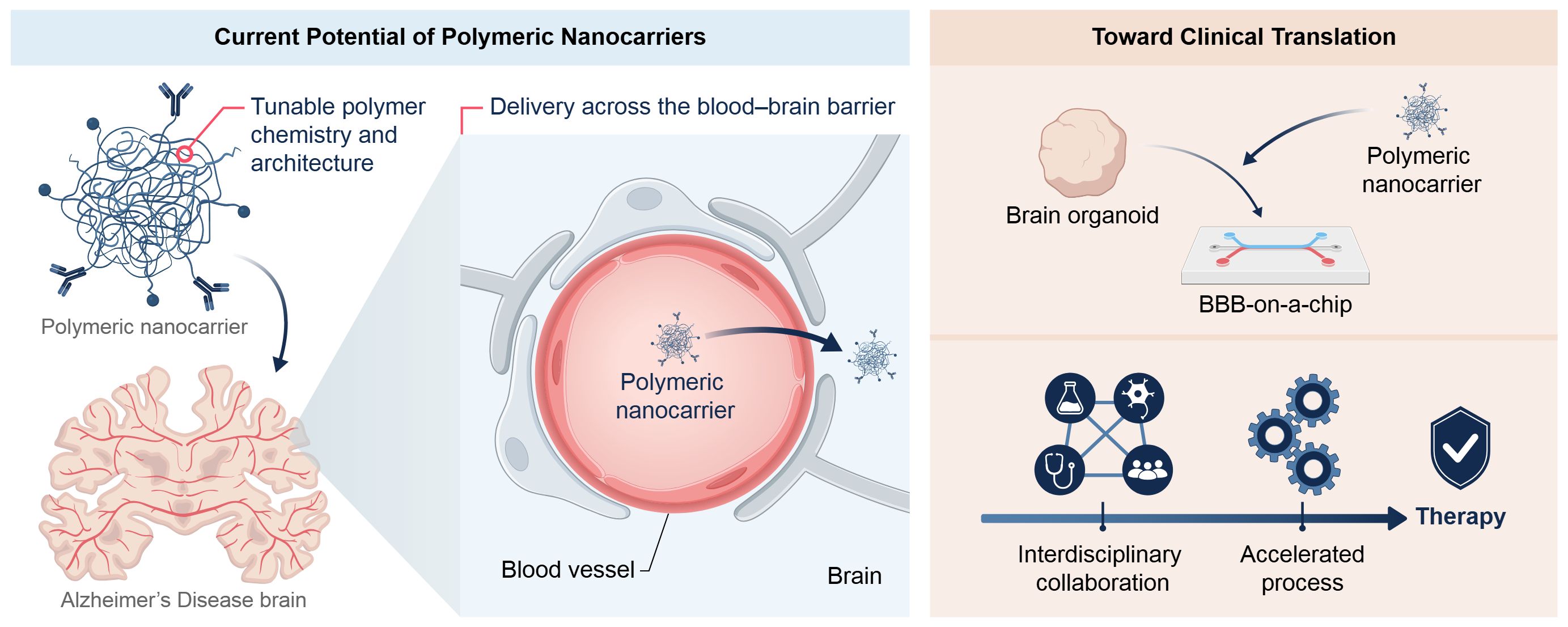
Synthetic polymer-based nanoparticle platforms are expected to play an increasingly important role in Alzheimer’s disease research and therapy, provided that future development prioritizes biological relevance, translational feasibility, and regulatory alignment. Despite substantial progress in carrier engineering, the safe and effective delivery of therapeutics across the blood–brain barrier and within the complex central nervous system microenvironment remains the principal challenge. Continued optimization of hybrid polymer architectures, surface functionalization strategies, and degradation-controlled systems will be essential to balance therapeutic efficacy with long-term biocompatibility. An important direction for the field is the integration of polymeric nanocarriers with advanced human-relevant models, including brain organoids, blood–brain barrier-on-a-chip platforms, and multi-organ microphysiological systems (Figure 4). These models offer more predictive assessment of nanoparticle transport, toxicity, and therapeutic response than conventional animal models, and are expected to play a key role in both preclinical optimization and regulatory-relevant validation. From a broader perspective, synthetic polymer-based nanoparticles have demonstrated considerable versatility across a range of biomedical applications, including oncology and vaccine and antigen delivery. However, neurodegenerative diseases present distinct biological and translational constraints, particularly with respect to brain access and chronic safety, underscoring the importance of maintaining a disease-focused development strategy. In this context, future progress in Alzheimer’s nanomedicine will depend on reducing formulation complexity, ensuring scalable manufacturing processes, and integrating regulatory considerations early in the design and development process. The next phase of polymeric nanomedicine for Alzheimer’s disease is likely to be shaped by hybrid material systems validated in advanced human models, alongside practical considerations of manufacturability and regulatory compliance. Strategic integration of materials science, neurobiology, and regulatory science will be essential for translating promising nanomedicine-based drug delivery concepts into clinically viable therapeutic strategies.
Synthetic polymeric nanoparticles hold significant potential to improve AD treatment. By encapsulating therapeutic agents, these nanoparticles can protect them from degradation, prolong systemic circulation, and importantly, facilitate their transport across the blood–brain barrier (BBB). The tunability of polymer chemistry and architecture enables the rational design of drug delivery systems capable of sustained release, targeted delivery, and controlled degradation, directly addressing key limitations of current AD treatments. Despite this potential, translation from preclinical development to clinical application remains challenging. Future efforts should focus on the development of next-generation polymeric nanocarriers with improved targeting specificity, stimuli-responsive release behavior, and enhanced long-term safety profiles. Increasing emphasis is also expected to be placed on validation using advanced human-relevant models, such as brain organoids and blood–brain barrier (BBB)-on-a-chip platforms, to more accurately model human physiology and support regulatory decision-making. Future advances in polymeric nanomedicine for Alzheimer’s disease will depend on close collaboration among materials scientists, neurobiologists, clinicians, and regulatory stakeholders. Equally important will be early consideration of manufacturability, scalability, and regulatory alignment to ensure clinical feasibility. Although polymer-based nanoparticles have demonstrated considerable versatility across a wide range of biomedical applications, their successful translation in Alzheimer’s disease will require disease-specific design strategies that address the unique biological and safety constraints of the central nervous system. Although the path forward remains complex, continued interdisciplinary integration and technological refinement provide a realistic pathway toward clinically meaningful therapies for Alzheimer’s disease (AD).
AD
Alzheimer’s Disease
APP
Amyloid Precursor Protein
Aβ
amyloid-β
BACE1
β-site Amyloid Precursor Protein Cleaving Enzyme 1
BBB
Blood–Brain Barrier
BBB-on-a-chip
Blood–Brain Barrier-on-a-chip
BIN1
Bridging Integrator 1
CLU
Clusterin
CMT
Carrier-Mediated Transport
CNS
Central Nervous System
EPR
Enhanced Permeability and Retention
FAD
Familial Alzheimer’s Disease
FDA
U.S. Food and Drug Administration
GMP
Good Manufacturing Practice
hPSC
Human Pluripotent Stem Cell
LMW
Low Molecular Weight
LRP1
Low-Density Lipoprotein Receptor–Related Protein 1
MEM@ PCL
Memantine-Loaded Poly(ε-caprolactone) nanocapsules
MENA
Middle East and North Africa
NAMs
New Approach Methodologies
NFT
Neurofibrillary Tangle
NMDA
N-Methyl-D-Aspartate
NP
Nanoparticle
PAMAM
Polyamidoamine
PCL
Poly(ε-caprolactone)
PEG
Poly(ethylene glycol)
PEI
Polyethylenimine
PLA
Poly(lactic acid)
PLGA
Poly(lactic-co-glycolic acid)
PSEN
Presenilin
RMT
Receptor-Mediated Transcytosis
SAD
Sporadic Alzheimer’s Disease
siRNA
Small Interfering RNA
TEER
Trans-Endothelial Electrical Resistance
TGN
TGNYKALHPHN Peptide
UAE
United Arab Emirates
Conceptualization: B.A., N.A., J.A., J.F.C., Z.M., and K.-i.K.; Methodology: B.A., N.A., J.A., J.F.C., Z.M., and K.-i.K.; Investigation: B.A., N.A., J.A., J.F.C., Z.M., and K.-i.K.; Writing—Original Draft Preparation: B.A., N.A., J.A., J.F.C., Z.M., and K.-i.K.; Writing—Review & Editing: B.A., N.A., J.A., J.F.C., Z.M., and K.-i.K.; Visualization: B.A., N.A., J.A., J.F.C., Z.M., and K.-i.K.; Supervision: K.-i.K.; Project Administration: K.-i.K. All authors have read and agreed to the published version of the manuscript.
The authors declare no conflicts of interest.
The study did not receive any external funding and was conducted using only institutional resources.
The authors acknowledge insightful discussions with colleagues. Figure 4 and the graphical abstract were prepared by Duygu Koldere Vilain (Life Science Editors Inc.).
ChatGPT-5.2 and Gemini 2.5 were used to improve English grammar and enhance readability.
[1] Karran, E.; De Strooper, B. The Amyloid Hypothesis in Alzheimer Disease: New Insights from New Therapeutics. Nat. Rev. Drug Discov. 2022, 21, 306–318. [CrossRef]
[2] Li, X.; Feng, X.; Sun, X.; Hou, N.; Han, F.; Liu, Y. Global, Regional, and National Burden of Alzheimer’s Disease and Other Dementias, 1990–2019. Front. Aging Neurosci. 2022, 14, 937486. [CrossRef]
[3] Safiri, S.; Noori, M.; Nejadghaderi, S.A.; Mousavi, S.E.; Sullman, M.J.M.; Araj-Khodaei, M.; Collins, G.S.; Kolahi, A.A.; Gharagozli, K. The Burden of Alzheimer’s Disease and Other Types of Dementia in the Middle East and North Africa Region, 1990–2019. Age Ageing 2023, 52, afad042. [CrossRef]
[4] Friedrich, R.P.; Tepper, K.; Rönicke, R.; Soom, M.; Westermann, M.; Reymann, K.; Kaether, C.; Fändrich, M. Mechanism of Amyloid Plaque Formation Suggests an Intracellular Basis of Aβ Pathogenicity. Proc. Natl. Acad. Sci. USA 2010, 107, 1942–1947. [CrossRef]
[5] Gu, D.; Ou, S.; Liu, G. Traumatic Brain Injury and Risk of Dementia and Alzheimer’s Disease: A Systematic Review and Meta-Analysis. Neuroepidemiology 2022, 56, 4–16. [CrossRef]
[6] Weaver, C. Conformational Change as One of the Earliest Alterations of Tau in Alzheimer’s Disease. Neurobiol. Aging 2000, 21, 719–727. [CrossRef] [PubMed]
[7] Graff-Radford, J.; Yong, K.X.X.; Apostolova, L.G.; Bouwman, F.H.; Carrillo, M.; Dickerson, B.C.; Rabinovici, G.D.; Schott, J.M.; Jones, D.T.; Murray, M.E. New Insights into Atypical Alzheimer’s Disease in the Era of Biomarkers. Lancet Neurol. 2021, 20, 222–234. [CrossRef]
[8] Serrano-Pozo, A.; Das, S.; Hyman, B.T. APOE and Alzheimer’s Disease: Advances in Genetics, Pathophysiology, and Therapeutic Approaches. Lancet Neurol. 2021, 20, 68–80. [CrossRef] [PubMed]
[9] Ionescu-Tucker, A.; Cotman, C.W. Emerging Roles of Oxidative Stress in Brain Aging and Alzheimer’s Disease. Neurobiol. Aging 2021, 107, 86–95. [CrossRef]
[10] Hampel, H.; Hardy, J.; Blennow, K.; Chen, C.; Perry, G.; Kim, S.H.; Villemagne, V.L.; Aisen, P.; Vendruscolo, M.; Iwatsubo, T.; et al. The Amyloid-β Pathway in Alzheimer’s Disease. Mol. Psychiatry 2021, 26, 5481–5503. [CrossRef] [PubMed]
[11] Sharma, K. Cholinesterase Inhibitors as Alzheimer’s Therapeutics (Review). Mol. Med. Rep. 2019, 20, 1479–1487. [CrossRef]
[12] Cummings, J.; Lee, G.; Ritter, A.; Sabbagh, M.; Zhong, K. Alzheimer’s Disease Drug Development Pipeline: 2020. Alzheimers Dement. Transl. Res. Clin. Interv. 2020, 6, e12050. [CrossRef]
[13] Birks, J.S.; Chong, L.Y.; Grimley Evans, J. Rivastigmine for Alzheimer’s Disease. Cochrane Database Syst. Rev. 2015, 9, CD001191. [CrossRef] [PubMed]
[14] Parsons, C.G.; Danysz, W.; Dekundy, A.; Pulte, I. Memantine and Cholinesterase Inhibitors: Complementary Mechanisms in the Treatment of Alzheimer’s Disease. Neurotox. Res. 2013, 24, 358–369. [CrossRef]
[15] Song, N.; Sun, S.; Chen, K.; Wang, Y.; Wang, H.; Meng, J.; Guo, M.; Zhang, X.D.; Zhang, R. Emerging Nanotechnology for Alzheimer’s Disease: From Detection to Treatment. J. Controlled Release 2023, 360, 392–417. [CrossRef]
[16] Wu, D.; Chen, Q.; Chen, X.; Han, F.; Chen, Z.; Wang, Y. The Blood–Brain Barrier: Structure, Regulation and Drug Delivery. Signal Transduct. Target. Ther. 2023, 8, 217. [CrossRef] [PubMed]
[17] Sweeney, M.D.; Sagare, A.P.; Zlokovic, B.V. Blood–Brain Barrier Breakdown in Alzheimer Disease and Other Neurodegenerative Disorders. Nat. Rev. Neurol. 2018, 14, 133–150. [CrossRef]
[18] Kadry, H.; Noorani, B.; Cucullo, L. A Blood–Brain Barrier Overview on Structure, Function, Impairment, and Biomarkers of Integrity. Fluids Barriers CNS 2020, 17, 69. [CrossRef]
[19] Harilal, S.; Jose, J.; Parambi, D.G.T.; Kumar, R.; Mathew, G.E.; Uddin, M.S.; Kim, H.; Mathew, B. Advancements in Nanotherapeutics for Alzheimer’s Disease: Current Perspectives. J. Pharm. Pharmacol. 2019, 71, 1370–1383. [CrossRef]
[20] Huang, Q.; Chen, Y.; Zhang, W.; Xia, X.; Li, H.; Qin, M.; Gao, H. Nanotechnology for Enhanced Nose-to-Brain Drug Delivery in Treating Neurological Diseases. J. Controlled Release 2024, 366, 519–534. [CrossRef] [PubMed]
[21] Ale, Y.; Nainwal, N. Progress and Challenges in the Diagnosis and Treatment of Brain Cancer Using Nanotechnology. Mol. Pharm. 2023, 20, 4893–4921. [CrossRef]
[22] Tang, W.; Fan, W.; Lau, J.; Deng, L.; Shen, Z.; Chen, X. Emerging Blood–Brain-Barrier-Crossing Nanotechnology for Brain Cancer Theranostics. Chem. Soc. Rev. 2019, 48, 2967–3014. [CrossRef]
[23] Di Mascolo, D.; Palange, A.L.; Primavera, R.; Macchi, F.; Catelani, T.; Piccardi, F.; Spanò, R.; Ferreira, M.; Marotta, R.; Armirotti, A.; et al. Conformable Hierarchically Engineered Polymeric Micromeshes Enabling Combinatorial Therapies in Brain Tumours. Nat. Nanotechnol. 2021, 16, 820–829. [CrossRef]
[24] Zheng, M.; Tao, W.; Zou, Y.; Farokhzad, O.C.; Shi, B. Nanotechnology-Based Strategies for siRNA Brain Delivery for Disease Therapy. Trends Biotechnol. 2018, 36, 562–575. [CrossRef] [PubMed]
[25] Sun, M.; Qin, F.; Bu, Q.; Zhao, Y.; Yang, X.; Zhang, D.; Cen, X. Exosome-Based Therapeutics: A Natural Solution to Overcoming the Blood–Brain Barrier in Neurodegenerative Diseases. MedComm 2025, 6, e70386. [CrossRef]
[26] Peng, H.; Ji, W.; Zhao, R.; Yang, J.; Lu, Z.; Li, Y.; Zhang, X. Exosome: A Significant Nano-Scale Drug Delivery Carrier. J. Mater. Chem. B 2020, 8, 7591–7608. [CrossRef]
[27] Jia, H.R.; Zhu, Y.X.; Duan, Q.Y.; Chen, Z.; Wu, F.G. Nanomaterials Meet Zebrafish: Toxicity Evaluation and Drug Delivery Applications. J. Controlled Release 2019, 311–312, 301–318. [CrossRef]
[28] Soetaert, F.; Korangath, P.; Serantes, D.; Fiering, S.; Ivkov, R. Cancer Therapy with Iron Oxide Nanoparticles: Agents of Thermal and Immune Therapies. Adv. Drug Deliv. Rev. 2020, 163–164, 65–83. [CrossRef] [PubMed]
[29] Zhang, W.; Mehta, A.; Tong, Z.; Esser, L.; Voelcker, N.H. Development of Polymeric Nanoparticles for Blood–Brain Barrier Transfer—Strategies and Challenges. Adv. Sci. 2021, 8, 2003937. [CrossRef]
[30] Matsumura, Y.; Maeda, H. A New Concept for Macromolecular Therapeutics in Cancer Chemotherapy: Mechanism of Tumoritropic Accumulation of Proteins and the Antitumor Agent Smancs. Cancer Res. 1986, 46, 6387–6392. [PubMed]
[31] Zhang, G.; Fu, X.; Sun, H.; Zhang, P.; Zhai, S.; Hao, J.; Cui, J.; Hu, M. Poly(Ethylene Glycol)-Mediated Assembly of Vaccine Particles to Improve Stability and Immunogenicity. ACS Appl. Mater. Interfaces 2021, 13, 13978–13989. [CrossRef]
[32] Knop, K.; Hoogenboom, R.; Fischer, D.; Schubert, U.S. Poly(Ethylene Glycol) in Drug Delivery: Pros and Cons as Well as Potential Alternatives. Angew. Chem. Int. Ed. 2010, 49, 6288–6308. [CrossRef]
[33] Barenholz, Y(Chezy). Doxil®—The First FDA-Approved Nano-Drug: Lessons Learned. J. Controlled Release 2012, 160, 117–134. [CrossRef]
[34] Zhang, Y.; Chen, H.; Li, R.; Sterling, K.; Song, W. Amyloid β-Based Therapy for Alzheimer’s Disease: Challenges, Successes and Future. Signal Transduct. Target. Ther. 2023, 8, 248. [CrossRef] [PubMed]
[35] Kim, J.; Um, H.; Kim, N.H.; Kim, D. Potential Alzheimer’s Disease Therapeutic Nano-Platform: Discovery of Amyloid-Beta Plaque Disaggregating Agent and Brain-Targeted Delivery System Using Porous Silicon Nanoparticles. Bioact. Mater. 2023, 24, 497–506. [CrossRef] [PubMed]
[36] Le Droumaguet, B.; Nicolas, J.; Brambilla, D.; Mura, S.; Maksimenko, A.; De Kimpe, L.; Salvati, E.; Zona, C.; Airoldi, C.; Canovi, M.; et al. Versatile and Efficient Targeting Using a Single Nanoparticulate Platform: Application to Cancer and Alzheimer’s Disease. ACS Nano 2012, 6, 5866–5879. [CrossRef]
[37] Baysal, I.; Yabanoglu-Ciftci, S.; Tunc-Sarisozen, Y.; Ulubayram, K.; Ucar, G. Interaction of Selegiline-Loaded PLGA-b-PEG Nanoparticles with Beta-Amyloid Fibrils. J. Neural Transm. 2013, 120, 903–910. [CrossRef]
[38] Wadhwa, G.; Venkata Krishna, K.; Kumar Dubey, S.; Taliyan, R. Design and Biological Evaluation of Repaglinide Loaded Polymeric Nanocarriers for Diabetes Linked Neurodegenerative Disorder: QbD-Driven Optimization, in Situ, in Vitro and in Vivo Investigation. Int. J. Pharm. 2023, 636, 122824. [CrossRef]
[39] Pisani, F.; Castagnola, V.; Simone, L.; Loiacono, F.; Svelto, M.; Benfenati, F. Role of Pericytes in Blood–Brain Barrier Preservation during Ischemia through Tunneling Nanotubes. Cell Death Dis. 2022, 13, 582. [CrossRef]
[40] Banks, W.A. Characteristics of Compounds That Cross the Blood–Brain Barrier. BMC Neurol. 2009, 9, S3. [CrossRef] [PubMed]
[41] Manek, E.; Petroianu, G.A. Brain Delivery of Antidotes by Polymeric Nanoparticles. J. Appl. Toxicol. 2021, 41, 20–32. [CrossRef] [PubMed]
[42] McLoughlin, C.D.; Nevins, S.; Stein, J.B.; Khakbiz, M.; Lee, K. Overcoming the Blood–Brain Barrier: Multifunctional Nanomaterial-Based Strategies for Targeted Drug Delivery in Neurological Disorders. Small Sci. 2024, 4, 2400232. [CrossRef]
[43] Ding, S.; Khan, A.I.; Cai, X.; Song, Y.; Lyu, Z.; Du, D.; Dutta, P.; Lin, Y. Overcoming Blood–Brain Barrier Transport: Advances in Nanoparticle-Based Drug Delivery Strategies. Mater. Today 2020, 37, 112–125. [CrossRef] [PubMed]
[44] Lu, Q.; Cai, X.; Zhang, X.; Li, S.; Song, Y.; Du, D.; Dutta, P.; Lin, Y. Synthetic Polymer Nanoparticles Functionalized with Different Ligands for Receptor-Mediated Transcytosis across the Blood–Brain Barrier. ACS Appl. Bio Mater. 2018, 1, 1687–1694. [CrossRef]
[45] Johnsen, K.B.; Moos, T. Revisiting Nanoparticle Technology for Blood–Brain Barrier Transport: Unfolding at the Endothelial Gate Improves the Fate of Transferrin Receptor-Targeted Liposomes. J. Controlled Release 2016, 222, 32–46. [CrossRef]
[46] Zhu, X.; Jin, K.; Huang, Y.; Pang, Z. Brain Drug Delivery by Adsorption-Mediated Transcytosis. In Brain Targeted Drug Delivery System; Elsevier, 2019; pp. 159–183. [CrossRef]
[47] La Barbera, L.; Mauri, E.; D’Amelio, M.; Gori, M. Functionalization Strategies of Polymeric Nanoparticles for Drug Delivery in Alzheimer’s Disease: Current Trends and Future Perspectives. Front. Neurosci. 2022, 16, 939855. [CrossRef]
[48] Demeule, M.; Currie, J.; Bertrand, Y.; Ché, C.; Nguyen, T.; Régina, A.; Gabathuler, R.; Castaigne, J.; Béliveau, R. Involvement of the Low-density Lipoprotein Receptor-related Protein in the Transcytosis of the Brain Delivery Vector Angiopep-2. J. Neurochem. 2008, 106, 1534–1544. [CrossRef]
[49] Al Jayoush, A.R.; Hassan, H.A.F.M.; Asiri, H.; Jafar, M.; Saeed, R.; Harati, R.; Haider, M. Niosomes for Nose-to-Brain Delivery: A Non-Invasive Versatile Carrier System for Drug Delivery in Neurodegenerative Diseases. J. Drug Deliv. Sci. Technol. 2023, 89, 105007. [CrossRef]
[50] Wang, X.; Yin, Y.; Zhou, H.; Chi, B.; Guan, L.; Li, P.; Li, J.; Wang, Y. Drug Delivery Pathways to the Central Nervous System via the Brain Glymphatic System Circumventing the Blood–brain Barrier. Exploration 2024, 5, 20240036. [CrossRef]
[51] Mura, S.; Nicolas, J.; Couvreur, P. Stimuli-Responsive Nanocarriers for Drug Delivery. Nat. Mater. 2013, 12, 991–1003. [CrossRef] [PubMed]
[52] Ensign, L.M.; Cone, R.; Hanes, J. Oral Drug Delivery with Polymeric Nanoparticles: The Gastrointestinal Mucus Barriers. Adv. Drug Deliv. Rev. 2012, 64, 557–570. [CrossRef] [PubMed]
[53] Saraiva, C.; Praça, C.; Ferreira, R.; Santos, T.; Ferreira, L.; Bernardino, L. Nanoparticle-Mediated Brain Drug Delivery: Overcoming Blood–Brain Barrier to Treat Neurodegenerative Diseases. J. Controlled Release 2016, 235, 34–47. [CrossRef]
[54] Sempf, K.; Arrey, T.; Gelperina, S.; Schorge, T.; Meyer, B.; Karas, M.; Kreuter, J. Adsorption of Plasma Proteins on Uncoated PLGA Nanoparticles. Eur. J. Pharm. Biopharm. 2013, 85, 53–60. [CrossRef]
[55] Liu, Q.; Xue, J.; Zhang, X.; Chai, J.; Qin, L.; Guan, J.; Zhang, X.; Mao, S. The Influence of a Biomimetic Pulmonary Surfactant Modification on the in Vivo Fate of Nanoparticles in the Lung. Acta Biomater. 2022, 147, 391–402. [CrossRef]
[56] Hühn, D.; Kantner, K.; Geidel, C.; Brandholt, S.; De Cock, I.; Soenen, S.J.H.; Rivera_Gil, P.; Montenegro, J.M.; Braeckmans, K.; Müllen, K.; et al. Polymer-Coated Nanoparticles Interacting with Proteins and Cells: Focusing on the Sign of the Net Charge. ACS Nano 2013, 7, 3253–3263. [CrossRef] [PubMed]
[57] Zhu, Y.; Xu, H.; Yu, C.; Jiang, W.; Hou, X.; Ma, M.; Wu, J. Polymers for the Treatment of Alzheimer’s Disease. Front. Pharmacol. 2025, 16, 1512941. [CrossRef]
[58] Hu, L.; Tao, Y.; Jiang, Y.; Qin, F. Recent Progress of Nanomedicine in the Treatment of Alzheimer’s Disease. Front. Cell Dev. Biol. 2023, 11, 1228679. [CrossRef]
[59] Wong, X.Y.; Sena-Torralba, A.; Álvarez-Diduk, R.; Muthoosamy, K.; Merkoçi, A. Nanomaterials for Nanotheranostics: Tuning Their Properties According to Disease Needs. ACS Nano 2020, 14, 2585–2627. [CrossRef]
[60] Agrawal, A.; Rellegadla, S.; Jain, S. Biomedical Applications of PLGA Particles. In Materials for Biomedical Engineering; Elsevier: Amsterdam, The Netherlands, 2019; pp. 87–129. [CrossRef]
[61] Liao, R.; Pon, J.; Chungyoun, M.; Nance, E. Enzymatic Protection and Biocompatibility Screening of Enzyme-Loaded Polymeric Nanoparticles for Neurotherapeutic Applications. Biomaterials 2020, 257, 120238. [CrossRef]
[62] Paul, P.S.; Cho, J.Y.; Wu, Q.; Karthivashan, G.; Grabovac, E.; Wille, H.; Kulka, M.; Kar, S. Unconjugated PLGA Nanoparticles Attenuate Temperature-Dependent β-Amyloid Aggregation and Protect Neurons against Toxicity: Implications for Alzheimer’s Disease Pathology. J. Nanobiotechnol. 2022, 20, 67. [CrossRef] [PubMed]
[63] Lee, L.Y.; Ranganath, S.H.; Fu, Y.; Zheng, J.L.; Lee, H.S.; Wang, C.H.; Smith, K.A. Paclitaxel Release from Micro-Porous PLGA Disks. Chem. Eng. Sci. 2009, 64, 4341–4349. [CrossRef]
[64] Makadia, H.K.; Siegel, S.J. Poly Lactic-Co-Glycolic Acid (PLGA) as Biodegradable Controlled Drug Delivery Carrier. Polymers 2011, 3, 1377–1397. [CrossRef]
[65] Bouissou, C.; Rouse, J.J.; Price, R.; Van Der Walle, C.F. The Influence of Surfactant on PLGA Microsphere Glass Transition and Water Sorption: Remodeling the Surface Morphology to Attenuate the Burst Release. Pharm. Res. 2006, 23, 1295–1305. [CrossRef]
[66] Jain, A.; Kunduru, K.R.; Basu, A.; Mizrahi, B.; Domb, A.J.; Khan, W. Injectable Formulations of Poly(Lactic Acid) and Its Copolymers in Clinical Use. Adv. Drug Deliv. Rev. 2016, 107, 213–227. [CrossRef]
[67] Danhier, F.; Ansorena, E.; Silva, J.M.; Coco, R.; Le Breton, A.; Préat, V. PLGA-Based Nanoparticles: An Overview of Biomedical Applications. J. Controlled Release 2012, 161, 505–522. [CrossRef] [PubMed]
[68] Alonso-Sande, M.; des Rieux, A.; Fievez, V.; Sarmento, B.; Delgado, A.; Evora, C.; Remuñán-López, C.; Préat, V.; Alonso, M.J. Development of PLGA-Mannosamine Nanoparticles as Oral Protein Carriers. Biomacromolecules 2013, 14, 4046–4052. [CrossRef] [PubMed]
[69] Omelczuk, M.O.; McGinity, J.W. The Influence of Polymer Glass Transition Temperature and Molecular Weight on Drug Release from Tablets Containing Poly(DL-Lactic Acid). Pharm. Res. 1992, 9, 26–32. [CrossRef] [PubMed]
[70] Anand, B.; Wu, Q.; Nakhaei-Nejad, M.; Karthivashan, G.; Dorosh, L.; Amidian, S.; Dahal, A.; Li, X.; Stepanova, M.; Wille, H.; et al. Significance of Native PLGA Nanoparticles in the Treatment of Alzheimer’s Disease Pathology. Bioact. Mater. 2022, 17, 506–525. [CrossRef]
[71] Shin, H.J.; Kim, I.S.; Choi, S.G.; Lee, K.; Park, H.; Shin, J.; Kim, D.; Beom, J.; Yi, Y.Y.; Gupta, D.P.; et al. Rejuvenating Aged Microglia by P16ink4a-siRNA-Loaded Nanoparticles Increases Amyloid-β Clearance in Animal Models of Alzheimer’s Disease. Mol. Neurodegener. 2024, 19, 25. [CrossRef]
[72] Liu, L.; He, H.; Du, B.; He, Y. Nanoscale Drug Formulations for the Treatment of Alzheimer’s Disease Progression. RSC Adv. 2025, 15, 4031–4078. [CrossRef]
[73] Tracy, G.C.; Huang, K.Y.; Hong, Y.T.; Ding, S.; Noblet, H.A.; Lim, K.H.; Kim, E.C.; Chung, H.J.; Kong, H. Intracerebral Nanoparticle Transport Facilitated by Alzheimer Pathology and Age. Nano Lett. 2023, 23, 10971–10982. [CrossRef]
[74] Shen, J.; Yuan, W.; Badv, M.; Moshaverinia, A.; Weiss, P.S. Modified Poly(ε-Caprolactone) with Tunable Degradability and Improved Biofunctionality for Regenerative Medicine. ACS Mater. Au 2023, 3, 540–547. [CrossRef]
[75] Zhu, B.; Chen, Y.; Lu, W.; Zhang, Q.; Gao, S.; Sun, L.; Chen, S.; Hu, R. A Biodegradable Long-Term Contraceptive Implant with Steady Levonorgestrel Release Based on PLGA Microspheres Embedded in PCL-Coated Implant. J. Drug Deliv. Sci. Technol. 2022, 67, 102955. [CrossRef]
[76] Mahmoudi, M.; Saeidian, H.; Mirjafary, Z.; Mokhtari, J. Preparation and Characterization of Memantine Loaded Polycaprolactone Nanocapsules for Alzheimer’s Disease. J. Porous Mater. 2021, 28, 205–212. [CrossRef]
[77] Mahmoudi, M.; Saeidian, H.; Mirjafary, Z.; Mokhtari, J. Controlled Release 7-Methoxytacrine-Polycaprolactone Nanocapsules Drug-Delivery System for Alzheimer’s Disease Treatment: Synthesis and Physico-Chemical Characterization. J. Polym. Environ. 2022, 30, 2280–2290. [CrossRef]
[78] Müller, V.; Caldas, B.S.; Radovanovic, E.; Muniz, E.C. Development of Hot Melt Extruded Polycaprolactone (PCL) Matrices for an Oral Ultra-Long-Lasting Delivery of Galantamine for Alzheimer’s Disease Therapy. J. Drug Deliv. Sci. Technol. 2025, 104, 106464. [CrossRef]
[79] Gu, J.; Yan, C.; Yin, S.; Wu, H.; Liu, C.; Xue, A.; Lei, X.; Zhang, N.; Geng, F. Erythrocyte Membrane-Coated Nanocarriers Modified by TGN for Alzheimer’s Disease. J. Controlled Release 2024, 366, 448–459. [CrossRef]
[80] Saha, S.K.; Tsuji, H. Effects of Rapid Crystallization on Hydrolytic Degradation and Mechanical Properties of Poly(l-Lactide-Co-ε-Caprolactone). React. Funct. Polym. 2006, 66, 1362–1372. [CrossRef]
[81] Jeong, S.I.; Kim, B.S.; Lee, Y.M.; Ihn, K.J.; Kim, S.H.; Kim, Y.H. Morphology of Elastic Poly(l -Lactide-Co-ε-Caprolactone) Copolymers and in Vitro and in Vivo Degradation Behavior of Their Scaffolds. Biomacromolecules 2004, 5, 1303–1309. [CrossRef]
[82] Prabhath, A.; Vernekar, V.N.; Vasu, V.; Badon, M.; Avochinou, J.; Asandei, A.D.; Kumbar, S.G.; Weber, E.; Laurencin, C.T. Kinetic Degradation and Biocompatibility Evaluation of Polycaprolactone-Based Biologics Delivery Matrices for Regenerative Engineering of the Rotator Cuff. J. Biomed. Mater. Res. A 2021, 109, 2137–2153. [CrossRef] [PubMed]
[83] Zhang, W.; Yu, M.; Cao, Y.; Zhuang, Z.; Zhang, K.; Chen, D.; Liu, W.; Yin, J. An Anti-Bacterial Porous Shape Memory Self-Adaptive Stiffened Polymer for Alveolar Bone Regeneration after Tooth Extraction. Bioact. Mater. 2023, 21, 450–463. [CrossRef]
[84] Roberts, C.T.; Grunlan, M.A. Biodegradable Polyesters: Approaches to Increase Degradation Rates for Biomedical Applications. ACS Macro Lett. 2025, 14, 1221–1240. [CrossRef]
[85] Roberts, C.T.; Beck, S.K.; Prejean, C.M.; Graul, L.M.; Maitland, D.J.; Grunlan, M.A. Star-PCL Shape Memory Polymer (SMP) Scaffolds with Tunable Transition Temperatures for Enhanced Utility. J. Mater. Chem. B 2024, 12, 3694–3702. [CrossRef]
[86] Nevoralová, M.; Koutný, M.; Ujčić, A.; Starý, Z.; Šerá, J.; Vlková, H.; Šlouf, M.; Fortelný, I.; Kruliš, Z. Structure Characterization and Biodegradation Rate of Poly(ε-Caprolactone)/Starch Blends. Front. Mater. 2020, 7, 141. [CrossRef]
[87] Massadeh, S.; Alaamery, M.; Al-Qatanani, S.; Alarifi, S.; Bawazeer, S.; Alyafee, Y. Synthesis of Protein-Coated Biocompatible Methotrexate-Loaded PLA-PEG-PLA Nanoparticles for Breast Cancer Treatment. Nano Rev. Exp. 2016, 7, 31996. [CrossRef]
[88] Zhang, X.; Chau, L.Y.; Chan, H.W.; Weng, J.; Wong, K.W.; Chow, S.F.; Chow, A.H.L. Physical Stability and in Vivo Brain Delivery of Polymeric Ibuprofen Nanoparticles Fabricated by Flash Nanoprecipitation. Int. J. Pharm. 2021, 598, 120224. [CrossRef] [PubMed]
[89] Habib, S.; Singh, M. Angiopep-2-Modified Nanoparticles for Brain-Directed Delivery of Therapeutics: A Review. Polymers 2022, 14, 712. [CrossRef]
[90] Cinar, E.; Mutluay, S.U.; Baysal, I.; Gultekinoglu, M.; Ulubayram, K.; Yabanoglu Ciftci, S.; Tel, B.C.; Ucar, G. Donepezil-Loaded PLGA-b-PEG Nanoparticles Enhance the Learning and Memory Function of Beta-Amyloid Rat Model of Alzheimer’s Disease. Arch. Neuropsychiatry 2022, 59, 281–289. [CrossRef]
[91] Chen, Y.; Pan, C.; Xuan, A.; Xu, L.; Bao, G.; Liu, F.; Fang, J.; Long, D. Treatment Efficacy of NGF Nanoparticles Combining Neural Stem Cell Transplantation on Alzheimer’s Disease Model Rats. Med. Sci. Monit. 2015, 21, 3608–3615. [CrossRef] [PubMed]
[92] Boussif, O.; Lezoualc’h, F.; Zanta, M.A.; Mergny, M.D.; Scherman, D.; Demeneix, B.; Behr, J.P. A Versatile Vector for Gene and Oligonucleotide Transfer into Cells in Culture and in Vivo: Polyethylenimine. Proc. Natl. Acad. Sci. USA 1995, 92, 7297–7301. [CrossRef]
[93] Ewe, A.; Höbel, S.; Heine, C.; Merz, L.; Kallendrusch, S.; Bechmann, I.; Merz, F.; Franke, H.; Aigner, A. Optimized Polyethylenimine (PEI)-Based Nanoparticles for siRNA Delivery, Analyzed in Vitro and in an Ex Vivo Tumor Tissue Slice Culture Model. Drug Deliv. Transl. Res. 2017, 7, 206–216. [CrossRef]
[94] Reinhard, S.; Wang, Y.; Dengler, S.; Wagner, E. Precise Enzymatic Cleavage Sites for Improved Bioactivity of siRNA Lipo-Polyplexes. Bioconjug. Chem. 2018, 29, 3649–3657. [CrossRef]
[95] Bonner, D.K.; Zhao, X.; Buss, H.; Langer, R.; Hammond, P.T. Crosslinked Linear Polyethylenimine Enhances Delivery of DNA to the Cytoplasm. J. Controlled Release 2013, 167, 101–107. [CrossRef]
[96] Aso, E.; Martinsson, I.; Appelhans, D.; Effenberg, C.; Benseny-Cases, N.; Cladera, J.; Gouras, G.; Ferrer, I.; Klementieva, O. Poly(Propylene Imine) Dendrimers with Histidine-Maltose Shell as Novel Type of Nanoparticles for Synapse and Memory Protection. Nanomedicine Nanotechnol. Biol. Med. 2019, 17, 198–209. [CrossRef]
[97] Benjaminsen, R.V.; Mattebjerg, M.A.; Henriksen, J.R.; Moghimi, S.M.; Andresen, T.L. The Possible “Proton Sponge” Effect of Polyethylenimine (PEI) Does Not Include Change in Lysosomal pH. Mol. Ther. 2013, 21, 149–157. [CrossRef]
[98] Akinc, A.; Thomas, M.; Klibanov, A.M.; Langer, R. Exploring Polyethylenimine-mediated DNA Transfection and the Proton Sponge Hypothesis. J. Gene Med. 2005, 7, 657–663. [CrossRef]
[99] Zhang, C.; Gu, Z.; Shen, L.; Liu, X.; Lin, H. A Dual Targeting Drug Delivery System for Penetrating Blood–Brain Barrier and Selectively Delivering siRNA to Neurons for Alzheimer’s Disease Treatment. Curr. Pharm. Biotechnol. 2017, 18, 1124–1131. [CrossRef]
[100] Godbey, W.T.; Wu, K.K.; Mikos, A.G. Poly(Ethylenimine)-Mediated Gene Delivery Affects Endothelial Cell Function and Viability. Biomaterials 2001, 22, 471–480. [CrossRef]
[101] Yang, C.; Cheng, W.; Teo, P.Y.; Engler, A.C.; Coady, D.J.; Hedrick, J.L.; Yang, Y.Y. Mitigated Cytotoxicity and Tremendously Enhanced Gene Transfection Efficiency of PEI through Facile One-Step Carbamate Modification. Adv. Healthc. Mater. 2013, 2, 1304–1308. [CrossRef]
[102] Ogris, M.; Brunner, S.; Schüller, S.; Kircheis, R.; Wagner, E. PEGylated DNA/Transferrin–PEI Complexes: Reduced Interaction with Blood Components, Extended Circulation in Blood and Potential for Systemic Gene Delivery. Gene Ther. 1999, 6, 595–605. [CrossRef]
[103] Petersen, H.; Fechner, P.M.; Fischer, D.; Kissel, T. Synthesis, Characterization, and Biocompatibility of Polyethylenimine- g Raft -Poly(Ethylene Glycol) Block Copolymers. Macromolecules 2002, 35, 6867–6874. [CrossRef]
[104] Gabrielson, N.P.; Pack, D.W. Acetylation of Polyethylenimine Enhances Gene Delivery via Weakened Polymer/DNA Interactions. Biomacromolecules 2006, 7, 2427–2435. [CrossRef]
[105] Mapfumo, P.P.; Reichel, L.S.; André, T.; Hoeppener, S.; Rudolph, L.K.; Traeger, A. Optimizing Biocompatibility and Gene Delivery with DMAEA and DMAEAm: A Niacin-Derived Copolymer Approach. Biomacromolecules 2024, 25, 4749–4761. [CrossRef]
[106] Erfani, A.; Seaberg, J.; Aichele, C.P.; Ramsey, J.D. Interactions between Biomolecules and Zwitterionic Moieties: A Review. Biomacromolecules 2020, 21, 2557–2573. [CrossRef]
[107] Sun, J.; Zeng, F.; Jian, H.; Wu, S. Grafting Zwitterionic Polymer Chains onto PEI as a Convenient Strategy to Enhance Gene Delivery Performance. Polym. Chem. 2013, 4, 5810. [CrossRef]
[108] Deng, R.; Yue, Y.; Jin, F.; Chen, Y.; Kung, H.F.; Lin, M.C.M.; Wu, C. Revisit the Complexation of PEI and DNA—How to Make Low Cytotoxic and Highly Efficient PEI Gene Transfection Non-Viral Vectors with a Controllable Chain Length and Structure? J. Controlled Release 2009, 140, 40–46. [CrossRef]
[109] Kamaly, N.; Yameen, B.; Wu, J.; Farokhzad, O.C. Degradable Controlled-Release Polymers and Polymeric Nanoparticles: Mechanisms of Controlling Drug Release. Chem. Rev. 2016, 116, 2602–2663. [CrossRef]
[110] Español, L.; Larrea, A.; Andreu, V.; Mendoza, G.; Arruebo, M.; Sebastian, V.; Aurora-Prado, M.S.; Kedor-Hackmann, E.R.M.; Santoro, M.I.R.M.; Santamaria, J. Dual Encapsulation of Hydrophobic and Hydrophilic Drugs in PLGA Nanoparticles by a Single-Step Method: Drug Delivery and Cytotoxicity Assays. RSC Adv. 2016, 6, 111060–111069. [CrossRef]
[111] Grossen, P.; Witzigmann, D.; Sieber, S.; Huwyler, J. PEG-PCL-Based Nanomedicines: A Biodegradable Drug Delivery System and Its Application. J. Controlled Release 2017, 260, 46–60. [CrossRef]
[112] Ravivarapu, H. Polymer and Microsphere Blending to Alter the Release of a Peptide from PLGA Microspheres. Eur. J. Pharm. Biopharm. 2000, 50, 263–270. [CrossRef]
[113] Sheikh Hasan, A.; Sapin, A.; Damgé, C.; Leroy, P.; Socha, M.; Maincent, P. Reduction of the in Vivo Burst Release of Insulin-Loaded Microparticles. J. Drug Deliv. Sci. Technol. 2015, 30, 486–493. [CrossRef]
[114] Sinha, V.R.; Bansal, K.; Kaushik, R.; Kumria, R.; Trehan, A. Poly-ϵ-Caprolactone Microspheres and Nanospheres: An Overview. Int. J. Pharm. 2004, 278, 1–23. [CrossRef]
[115] Otsuka, H.; Nagasaki, Y.; Kataoka, K. PEGylated Nanoparticles for Biological and Pharmaceutical Applications. Adv. Drug Deliv. Rev. 2003, 55, 403–419. [CrossRef]
[116] Casper, J.; Schenk, S.H.; Parhizkar, E.; Detampel, P.; Dehshahri, A.; Huwyler, J. Polyethylenimine (PEI) in Gene Therapy: Current Status and Clinical Applications. J. Controlled Release 2023, 362, 667–691. [CrossRef]
[117] Midoux, P.; Pichon, C.; Yaouanc, J.; Jaffrès, P. Chemical Vectors for Gene Delivery: A Current Review on Polymers, Peptides and Lipids Containing Histidine or Imidazole as Nucleic Acids Carriers. Br. J. Pharmacol. 2009, 157, 166–178. [CrossRef]
[118] Hersh, A.M.; Alomari, S.; Tyler, B.M. Crossing the Blood–Brain Barrier: Advances in Nanoparticle Technology for Drug Delivery in Neuro-Oncology. Int. J. Mol. Sci. 2022, 23, 4153. [CrossRef]
[119] Xie, J.; Shen, Z.; Anraku, Y.; Kataoka, K.; Chen, X. Nanomaterial-Based Blood–Brain-Barrier (BBB) Crossing Strategies. Biomaterials 2019, 224, 119491. [CrossRef]
[120] Liu, S.; Jin, X.; Ge, Y.; Dong, J.; Liu, X.; Pei, X.; Wang, P.; Wang, B.; Chang, Y.; Yu, X. Advances in Brain-Targeted Delivery Strategies and Natural Product-Mediated Enhancement of Blood–Brain Barrier Permeability. J. Nanobiotechnol. 2025, 23, 382. [CrossRef]
[121] Hervé, F.; Ghinea, N.; Scherrmann, J.M. CNS Delivery Via Adsorptive Transcytosis. AAPS J. 2008, 10, 455–472. [CrossRef]
[122] Lu, W.; Zhang, Y.; Tan, Y.Z.; Hu, K.L.; Jiang, X.G.; Fu, S.K. Cationic Albumin-Conjugated Pegylated Nanoparticles as Novel Drug Carrier for Brain Delivery. J. Controlled Release 2005, 107, 428–448. [CrossRef]
[123] Zolnik, B.S.; Burgess, D.J. Effect of Acidic pH on PLGA Microsphere Degradation and Release. J. Controlled Release 2007, 122, 338–344. [CrossRef]
[124] Ma, S.; Feng, X.; Liu, F.; Wang, B.; Zhang, H.; Niu, X. The Pro-inflammatory Response of Macrophages Regulated by Acid Degradation Products of Poly(Lactide-co-glycolide) Nanoparticles. Eng. Life Sci. 2021, 21, 709–720. [CrossRef]
[125] McSweeney, M.D.; Price, L.S.L.; Wessler, T.; Ciociola, E.C.; Herity, L.B.; Piscitelli, J.A.; DeWalle, A.C.; Harris, T.N.; Chan, A.K.P.; Saw, R.S.; et al. Overcoming Anti-PEG Antibody Mediated Accelerated Blood Clearance of PEGylated Liposomes by Pre-Infusion with High Molecular Weight Free PEG. J. Controlled Release 2019, 311–312, 138–146. [CrossRef]
[126] Chang, T.C.; Chen, B.M.; Wu, J.Y.; Cheng, T.L.; Roffler, S. Impact of Anti-PEG Antibody Affinity on Accelerated Blood Clearance of Pegylated Epoetin Beta in Mice. Biomed. Pharmacother. 2022, 146, 112502. [CrossRef]
[127] Gothwal, A.; Kumar, H.; Nakhate, K.T.; Ajazuddin; Dutta, A.; Borah, A.; Gupta, U. Lactoferrin Coupled Lower Generation PAMAM Dendrimers for Brain Targeted Delivery of Memantine in Aluminum-Chloride-Induced Alzheimer’s Disease in Mice. Bioconjug. Chem. 2019, 30, 2573–2583. [CrossRef]
[128] Fox, L.J.; Richardson, R.M.; Briscoe, W.H. PAMAM Dendrimer—Cell Membrane Interactions. Adv. Colloid Interface Sci. 2018, 257, 1–18. [CrossRef]
[129] Brenza, T.M.; Schlichtmann, B.W.; Bhargavan, B.; Vela Ramirez, J.E.; Nelson, R.D.; Panthani, M.G.; McMillan, J.M.; Kalyanaraman, B.; Gendelman, H.E.; Anantharam, V.; et al. Biodegradable Polyanhydride-based Nanomedicines for Blood to Brain Drug Delivery. J. Biomed. Mater. Res. A 2018, 106, 2881–2890. [CrossRef]
[130] Attenello, F.J.; Mukherjee, D.; Datoo, G.; McGirt, M.J.; Bohan, E.; Weingart, J.D.; Olivi, A.; Quinones-Hinojosa, A.; Brem, H. Use of Gliadel (BCNU) Wafer in the Surgical Treatment of Malignant Glioma: A 10-Year Institutional Experience. Ann. Surg. Oncol. 2008, 15, 2887–2893. [CrossRef]
[131] Commissioner, O. FDA Announces Plan to Phase Out Animal Testing Requirement for Monoclonal Antibodies and Other Drugs; FDA: Silver Spring, MD, USA, 2025 Available online:; Available online: https://www.fda.gov/news-events/press-announcements/fda-announces-plan-phase-out-animal-testing-requirement-monoclonal-antibodies-and-other-drugs (accessed on 9 January 2026).
[132] National Institutes of Health (NIH). NIH to Prioritize Human-Based Research Technologies. Available online: https://www.nih.gov/news-events/news-releases/nih-prioritize-human-based-research-technologies (accessed on 9 January 2026).
[133] Fernandes, S.; Revanna, J.; Pratt, J.; Hayes, N.; Marchetto, M.C.; Gage, F.H. Modeling Alzheimer’s Disease Using Human Cell Derived Brain Organoids and 3D Models. Front. Neurosci. 2024, 18, 1434945. [CrossRef]
[134] Cho, A.N.; Jin, Y.; An, Y.; Kim, J.; Choi, Y.S.; Lee, J.S.; Kim, J.; Choi, W.Y.; Koo, D.J.; Yu, W.; et al. Microfluidic Device with Brain Extracellular Matrix Promotes Structural and Functional Maturation of Human Brain Organoids. Nat. Commun. 2021, 12, 4730. [CrossRef]
[135] Tsai, Y.C. Proof of Concept for Brain Organoid-on-a-Chip to Create Multiple Domains in Forebrain Organoids. RSC Adv. 2025, 15, 3749–3755. [CrossRef]
[136] Choe, M.S.; Yeo, H.C.; Kim, J.S.; Lee, J.; Lee, H.J.; Kim, H.R.; Baek, K.M.; Jung, N.Y.; Choi, M.; Lee, M.Y. Simple Modeling of Familial Alzheimer’s Disease Using Human Pluripotent Stem Cell-Derived Cerebral Organoid Technology. Stem Cell Res. Ther. 2024, 15, 118. [CrossRef] [PubMed]
[137] Luo, J.; Li, P. Human Pluripotent Stem Cell-Derived Brain Organoids as in Vitro Models for Studying Neural Disorders and Cancer. Cell Biosci. 2021, 11, 99. [CrossRef]
[138] Sreenivasamurthy, S.; Laul, M.; Zhao, N.; Kim, T.; Zhu, D. Current Progress of Cerebral Organoids for Modeling Alzheimer’s Disease Origins and Mechanisms. Bioeng. Transl. Med. 2023, 8, e10378. [CrossRef]
[139] Da Silva, G.G.; Sacomani, D.P.; De Carvalho, B.G.; Porcionatto, M.A.; Gobbi, A.; Lima, R.S.; De La Torre, L.G. Microfluidic Systems to Mimic the Blood–Brain Barrier: From Market to Engineering Challenges and Perspectives. ACS Biomater. Sci. Eng. 2025, 11, 3789–3815. [CrossRef]
[140] Kantawala, B.; Shariff, S.; Ramadan, N.; Fawaz, V.; Hassan, Y.; Mugisha, N.; Yenkoyan, K.; Nazir, A.; Uwishema, O. Revolutionizing Neurotherapeutics: Blood–Brain Barrier-on-a-Chip Technologies for Precise Drug Delivery. Ann. Med. Surg. 2024, 86, 2794–2804. [CrossRef]
[141] Campisi, M.; Shin, Y.; Osaki, T.; Hajal, C.; Chiono, V.; Kamm, R.D. 3D Self-Organized Microvascular Model of the Human Blood–Brain Barrier with Endothelial Cells, Pericytes and Astrocytes. Biomaterials 2018, 180, 117–129. [CrossRef]
[142] Yoon, J.; Kim, J.; Shah, Z.; Awasthi, A.; Mahajan, A.; Kim, Y. Advanced Human BBB-on-a-Chip: A New Platform for Alzheimer’s Disease Studies. Adv. Healthc. Mater. 2021, 10, 2002285. [CrossRef]
[143] Ahn, S.I.; Sei, Y.J.; Park, H.J.; Kim, J.; Ryu, Y.; Choi, J.J.; Sung, H.J.; MacDonald, T.J.; Levey, A.I.; Kim, Y. Microengineered Human Blood–Brain Barrier Platform for Understanding Nanoparticle Transport Mechanisms. Nat. Commun. 2020, 11, 175. [CrossRef]
[144] Palma-Florez, S.; López-Canosa, A.; Moralez-Zavala, F.; Castaño, O.; Kogan, M.J.; Samitier, J.; Lagunas, A.; Mir, M. BBB-on-a-Chip with Integrated Micro-TEER for Permeability Evaluation of Multi-Functionalized Gold Nanorods against Alzheimer’s Disease. J. Nanobiotechnol. 2023, 21, 115. [CrossRef]
[145] Gawne, P.J.; Ferreira, M.; Papaluca, M.; Grimm, J.; Decuzzi, P. New Opportunities and Old Challenges in the Clinical Translation of Nanotheranostics. Nat. Rev. Mater. 2023, 8, 783–798. [CrossRef]
[146] Thapa, R.K.; Kim, J.O. Nanomedicine-Based Commercial Formulations: Current Developments and Future Prospects. J. Pharm. Investig. 2023, 53, 19–33. [CrossRef]
[147] DelveInsight. Drug Regulatory Affairs in the MENA | MENA Medicines Regulation|MENA. DelveInsight Business Research. Available online: https://www.delveinsight.com/blog/the-regulatory-process-for-drug-approval-in-the-mena-region (accessed on 9 January 2026).
[148] Pharmaceuticals and Medical Devices Agency. Nanomedicines. Available online: https://www.pmda.go.jp/english/review-services/regulatory-info/0011.html (accessed on 9 January 2026).
[149] Hertig, J.B.; Shah, V.P.; Flühmann, B.; Mühlebach, S.; Stemer, G.; Surugue, J.; Moss, R.; Di Francesco, T. Tackling the Challenges of Nanomedicines: Are We Ready? Am. J. Health Syst. Pharm. 2021, 78, 1047–1056. [CrossRef] [PubMed]
[150] Tutubala, T.E.; Egunlusi, A.O.; Fisher, D.; Dube, A.; Joubert, J. Nanomedicine Solutions for Alzheimer’s Disease: A Critical Review of Therapeutic Nanoparticle Strategies. ACS Omega 2025, 10, 53633–53657. [CrossRef] [PubMed]
Disclaimer/Publisher’s Note: The views expressed in this article are those of the author(s) and do not necessarily reflect the views of the publisher or editors. The publisher and editors assume no responsibility for any injury or damage resulting from the use of information contained herein.
©2026 Copyright by the Authors.
Licensed as an open access article distributed under the terms and conditions of the CC BY 4.0 license
We use cookies to improve your experience on our site. By continuing to use our site, you accept our use of cookies. Learn more