
 ACCESS
Research Article
ACCESS
Research Article
Volume 3, Article ID: 2026.0026

Vladimir Vasilyevich Glinkin
vvsyz1@gmail.com


Victoria Vladimirovna Glinkina
glinkinavic@yandex.ru

1 Federal State Budgetary Educational Institution of Higher Education "Donetsk State Medical University named after M. Gorky" of the Ministry of Health of the Russian Federation, Donetsk, Russia
2 State Budgetary Institution "Donetsk City Dental Clinic No. 1", Donetsk, Russia
* Author to whom correspondence should be addressed
Received: 05 Jun 2025 Accepted: 06 Mar 2026 Available Online: 07 Mar 2026
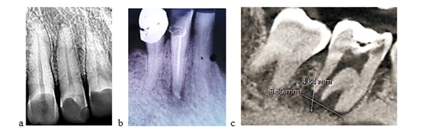
Introduction: High-quality root canal obturation and the correct choice of sealant are the main factors preventing its reinfection. A wide lumen of the apical foramen, often of inflammatory genesis, actually excludes the possibility of root canal sealing. Objective: to conduct, using SEM, morphological studies of endo-sealers used for filling canals of teeth with destructive forms of periodontitis and the quality of sealing of the root canal with destroyed apical constriction. Materials and methods: Scanning electron microscopy (SEM) methods were used to study the morphology and microstructure of dental samples and filling materials. To verify the size of the apical foramen, we developed a method using a gutta-percha pin. Three materials were used as endosealers: Foredent, Sealapex, Trioxident. To treat patients with partially and severely destroyed apical constriction, we proposed and introduced into practice a technique of orthograde root canal filling with Trioxident cement. Sections of 15 roots of extracted teeth were prepared and divided into 3 groups depending on the endo-sealant used. Results: The polymerization process of filling materials leads to shrinkage and the formation of micro cracks and micropores in them. Sealapex has an intermediate value of 7.32 µm (p <0.05). The average sizes of the gaps between the endosealant and the tooth tissue in the area of the apical foramen were: Foredent 27.13±3.58; Sealapex 66.81±3.57; Trioxident 6.51±3.55. Conclusions: The presence of microcracks and micropores in the studied materials can be explained by shrinkage during hardening. The average size of microcracks between the root canal wall and the endodontic filling material for Foredent was 16.9 µm, which is 15 times higher than the Trioxident values (1.11 µm). The presence of the hydration process in Trioxident cement allows us to assume that its use for orthograde root canal filling will prevent the resorption of the filling material in the apical part of the tooth root and prevent the progression of the pathological process in the periodontium.
Disclaimer : This is not the final version of the article. Changes may occur when the manuscript is published in its final format.