

APA Style
Vladimir N. Pak. (2026). Here, There, and Everywhere: Alpha-Fetoprotein in Cancer Immunotherapy. Cancer Research and Therapy Connect, 2 (Article ID: 0011). https://doi.org/10.69709/TCTC.2025.121204MLA Style
Vladimir N. Pak. "Here, There, and Everywhere: Alpha-Fetoprotein in Cancer Immunotherapy". Cancer Research and Therapy Connect, vol. 2, 2026, Article ID: 0011, https://doi.org/10.69709/TCTC.2025.121204.Chicago Style
Vladimir N. Pak. 2026. "Here, There, and Everywhere: Alpha-Fetoprotein in Cancer Immunotherapy." Cancer Research and Therapy Connect 2 (2026): 0011. https://doi.org/10.69709/TCTC.2025.121204.
ACCESS
Review Article
Volume 2, Article ID: 2026.0011

Vladimir N. Pak
oncoshut@gmail.com
Independent Researcher, Toronto, ON, Canada
Received: 02 Dec 2025 Accepted: 01 Feb 2026 Available Online: 02 Feb 2026 Published: 31 Mar 2026
Alpha-fetoprotein (AFP) delivers nutrients to immature cells in a shuttle-like manner via alpha-fetoprotein receptor (AFPR)-mediated endocytosis. A small subset of immature myeloid-derived suppressor cells (MDSCs) acts as a key regulator of immune tolerance during pregnancy, cancer, and other conditions. MDSCs, low doses of AFP, and AFP-binding ligands can modulate both the innate and adaptive immune responses. MDSCs decrease excessive immune activation, while their depletion can reverse immune suppression. Reduction of MDSCs using AFP–toxin combinations reactivate natural killer (NK) cells, macrophages, and cytotoxic lymphocytes (CTLs), thereby enhancing both innate and adaptive immune responses. AFP combined with apoptosis-inducing toxins specifically destroys MDSCs and cancer cells without generating pro-inflammatory byproducts. AFP-toxin complexes or chemical conjugates demonstrate high efficacy, low toxicity, a well-defined mechanism of action, cost-effectiveness, and do not require personalization. AFP combinations with drugs or traditional medicines represent a targeted immune/chemotherapy approach for cancer prevention and treatment.
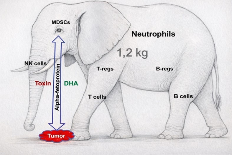
Cancer remains one of the leading causes of mortality, and many treatments aim to directly eliminate tumor cells. However, the immune system can eliminate such cells regularly. In cancer, the immune system is tolerant of the malignant cells. Reactivating the immune system is a physiological strategy that enables it to recognize and eliminate abnormal cells. The immunology of pregnancy and cancer is similar, with key cells employing mechanisms to evade immune attack [1,2]. Myeloid-derived suppressor cells (MDSCs) are immature myeloid progenitors released from the bone marrow or spleen during pregnancy and under chronic inflammatory conditions, such as cancer and other diseases. MDSCs include two major subsets based on their phenotypic and morphological features: monocytic MDSC (M-MDSC) and polymorphonuclear MDSC (PMN-MDSC, or former G-MDSC) [3]. Normally rare, these cells expand in cancer and exhibit suppressive functions that inhibit both innate and adaptive immunity [4]. MDSCs are essential for maternal–fetal tolerance during pregnancy [5,6], and are also co-opted by tumors. Their presence in cancer creates an immunosuppressive tumor microenvironment (TME) [7]. MDSCs inhibit natural killer (NK) cells [8]—and macrophage-mediated clearance of embryonic or tumor cells. They also promote the expansion of regulatory T cells (Tregs), thereby suppressing T- and B-cell responses. Over months to years of tumor development, an immune system composed of approximately 1.8 trillion cells (about 1.2 kg of immune biomass) [9] is unable to eradicate small populations of cancer cells once MDSCs impair immune recognition, effectively rendering the host immune system “blind” and preventing a functional antitumor response (Figure 1). Reflecting their central importance, more than 8600 PubMed-indexed publications now address MDSCs in cancer, yet only a small fraction investigate the interplay among MDSCs, oncofetal alpha-fetoprotein (AFP), and AFP-binding ligands. AFP supports fetal growth by transporting nutrients and promoting immune tolerance. In healthy adults, AFP expression is minimal; however, it reappears in several malignancies, most notably hepatocellular carcinoma (HCC) [10], germ cell tumors, and certain gastrointestinal cancers [11]. Elevated AFP often correlates with tumor burden, aggressiveness, and poor prognosis. Tumors exploit the same immunoregulatory pathways used during pregnancy to maintain tolerance to semi-allogeneic fetal tissue, thereby dampening immune responses [12]. AFP transports nutrients and drugs via the AFP receptor (AFPR), thereby modulating the activity of immune regulatory cells. AFPR is expressed on human T lymphocytes during blast transformation, as well as on human monocytes, primary macrophages, and cancer cells [13-16]. The AFPR structure has not yet been elucidated, and several other AFP-binding proteins have been identified, including chemokine receptors, mucins, and scavenger receptors, as well as metastasis-related and intracytoplasmic proteins [17,18]. This article focuses on AFPR-mediated endocytosis of AFP bound to toxins. When AFP selectively delivers toxins to AFPR+ regulatory immune and cancer cells, it supports cancer immunotherapy and cancer prevention [19]. Traditional medicine provides numerous bioactive compounds with cytotoxic, anti-inflammatory, and immune-modulating properties. Many of these compounds exhibit anticancer activity while also enhancing immune function [20,21]. When delivered by AFP, such compounds may gain selective access to MDSCs and tumor cells, potentially increasing efficacy while reducing systemic toxicity. This paper proposes a novel concept in immunotherapy. When delivering nutrients or drugs, AFP may suppress hyperactive immune responses, whereas AFP complexed with toxic compounds from traditional medicine may selectively deplete MDSCs, restore immune effector functions, and directly destroy tumor cells. This approach provides a universal, non-personalized, and low-toxicity strategy for cancer prevention and treatment. By modulating immune activity, this therapy enables the body’s own immune cells to recognize and eliminate malignant cells.
The structure, biochemical properties, and clinical roles of AFP have been thoroughly reviewed in the literature [22-27]. AFP is a 70 kDa oncofetal glycoprotein predominantly synthesized by the fetal liver, yolk sac, and gastrointestinal (GI) tract during embryonic development and is well recognized as an immunosuppressive protein [28]. AFP is used as a biomarker for pregnancy [29]. Elevated AFP levels correlate with pregnancy disorders and poor tumor prognosis [30]. AFP is a globular protein with 3–5% glycosylation and a flexible hydrophobic pocket that can accommodate fatty acids, bilirubin, steroids, xenobiotics, drugs, and other small molecules, enabling it to function as a natural shuttle carrier protein. During laboratory testing, the saturated palmitic (C16:0) and stearic (C18:0) fatty acids were extracted from 4 binding sites of AFP [31]. Naturally, AFP’s hydrophobic cavity fits 1–2 molecules of polyunsaturated fatty acids (PUFAs) like C22:6 docosahexaenoic acid (DHA) (Figure 2). Like the conformational change of hemoglobin upon oxygen binding, DHA binding alters AFP’s conformation [32], shifts its isoelectric point, enhances binding affinity, and stabilizes AFP: ligand complexes [33,34]. During its 3–5-day half-life, AFP naturally shuttles dozens of essential ligands into the embryo and other AFPR+ cells. Receptor-mediated endocytosis of AFP-ligand complexes occurs in the placenta, in cancer cells, human B-lymphoma and T-leukemia cells, and peripheral blood mononuclear cells (PBMC) [35-38]. The specific AFPR-mediated endocytosis by the small PBMC fraction—M-MDSCs (~1%)—was discovered by the following experiment. The AFP–daunorubicin conjugate eliminated ~60% of M-MDSCs, compared with ~8% cell death induced by daunorubicin alone. In contrast, G-MDSCs showed only minimal changes in viability (~18% versus ~20%). Notably, non-MDSC populations remained viable following treatments [39]. This discovery was particularly significant, expanding the relevance of AFP-based delivery to a major immunosuppressive cell population. AFP crosses the three cellular layers of the hemochorial placenta via AFPR, present in the normal human placenta [35], and/or the neonatal Fc receptor (FcRn) [40], returning with nutrients. AFP affinity for the essential PUFA DHA is 54 times stronger than that of albumin [41], and binds it even in a massive excess of albumin in the mother’s blood (35–55 mg/mL) compared to AFP (~150 ng/mL). Interestingly, AFP-DHA complexes remain stable even during chromatographic or electrophoretic procedures. The preferential binding of a ligand to AFP over albumin causes a significant enhancement of its fetal uptake. Thus, over 70% of estrone and estradiol injected into the maternal circulation of rats have been subsequently found to associate with AFP in the fetus. Unlike human AFP, rodent AFP binds strongly to these hormones, while artificial estrogens with low binding affinity do not concentrate in the rat fetus [42]. The AFP binding pocket can accommodate dioxin or diethylstilbestrol, providing a mechanistic explanation for their known embryotoxicity [43,44]. Nevertheless, as mutagens and carcinogens, these toxins cannot be used for cancer treatment. On the other hand, cyclophosphamide, doxorubicin, bleomycin, vincristine, and etoposide do not bind AFP strongly enough. They may be given safely to a woman in need during any trimester of pregnancy, as they do not harm the mother or fetus [45]. Porcine AFP (pAFP) shares extensive amino acid and functional homology with AFP (Figure 3). Unlike AFP, which has several glycosylated isoforms, mono-glycosylated pAFP serves both nutrient-delivery and immunosuppressive functions. PAFP binds ~2.6 moles of DHA and arachidonic acid per mole of protein [46]. Notably, pAFP transports nutrients and ligands across the six cellular layers of the epitheliochorial placenta, demonstrating exceptional transcytosis efficiency and evolutionary specialization for high-capacity ligand delivery [47]. AFP-bound cytotoxic compounds are selectively internalized by cancer cells through AFPR-mediated endocytosis. Electron microscopy has been used to follow AFP conjugates with horseradish peroxidase after specific endocytosis. AFP has been observed within coated pits of the plasma membrane and has been tracked to vesicles, endosomes, and a tubular-vesicular network localized in the Golgi–centrosome region adjacent to the nucleus [48]. Once inside the cell, toxins can destroy organelles and induce apoptosis, autodigestion, or other regulated forms of cell death. For example, AFP delivers the glycoside atractyloside (ATR) [49] into AFPR+ cells, where ATR induces mitochondrial damage and subsequent apoptosis, representing a point of no return (Figure 3) [50].
Physiological AFP concentrations—approximately 5–10 ng/mL in healthy adults and 10–150 ng/mL during pregnancy—help maintain immune tolerance. Moderately elevated AFP levels (>7 ng/mL) in otherwise healthy individuals have been associated with protective metabolic phenotypes, including reduced hepatic steatosis, myosteatosis, and sarcopenia [51]. AFP administration increased muscle strength and endurance in humans and mice; it enhanced the relative mass of immunotropic organs, improved survival in aged mice, and reduced their auto-aggressive behavior [52,53]. The effects of AFP on immune cells are summarized in Table 1 [54-62]. The AFP effects on immune cells. Fetal-derived AFP (4 µg/kg/day) produced complete clinical responses in inflammatory bowel disease (IBD), enabling mucosal healing and reducing steroid use [52]. Recombinant not glycosylated AFP (rAFP) was used in patients with active rheumatoid arthritis [63]. The newer rAFP formulation (ACT-101) surpassed anti-tumor necrosis factor alpha antibodies in preclinical colitis models and improved symptoms in myasthenia gravis and IBD [64-66]. AFP transports nutrients and modulates the immune response via immature myeloid cells. The AFP properties depend on its ligands [67]. Hence, MDSC is a “double-edged sword,” playing protective or pathological roles depending on the level of AFP delivery. This trio mediates immune protection in autoimmune diseases—including multiple sclerosis, rheumatoid arthritis, and IBD—as well as in allergic conditions and organ transplantation. On the other hand, they promote cancer progression [68,69]. When bound to cytotoxic ligands, AFP becomes a targeted delivery system that addresses MDSCs and tumor cells, enabling a dual therapeutic effect: a comprehensive approach to cancer immunotherapy and direct cytotoxicity [70].Immune Cell Type/Process
AFP Effect
Functional Outcome
References
Monocytes
Downregulates major histocompatibility complex class II (MHC II) expression
Reduced antigen presentation capacity
[54]
Macrophages
Promotes polarization toward an M2-like phenotype
Immunosuppressive, pro-tumor macrophage profile
[55]
NK cells
Suppression
Decreased NK-mediated cytotoxicity
[56,58]
DCs
Inhibits DC function
Suppression of NK cell cytotoxicity
[57]
Human mononuclear leukocytes
Modulates differentiation and functional activity
Broad immunoregulatory effects
[59]
Tregs
Inhibition
Decreased number
[60]
T helper cells
Influences conversion of naïve T helpers into memory T cells
Modulation of adaptive immune responses
[61]
MDSCs
Modulates differentiation and functional activity
Broad immunoregulatory effects
[62]
The pore-forming anti-fungal antibiotic amphotericin B (AmB) disrupts organelles’ membranes, while sparing the plasma membrane, leading to cellular autodigestion. Patients with stage IV malignancies were infused with AFP (75–300 µg), which can bind AmB in the blood. Infusions frequently triggered acute-phase reactions—transient chills and fever. Notably, no signs of endotoxicity associated with rapid tumor lysis were observed. By the end of treatment, three patients demonstrated 30–40% reductions in primary tumor mass and metastatic burden. Two patients with lung cancer experienced continued metastatic regression for up to three months post-therapy. A patient with cerebral metastases demonstrated marked neurological improvement, including recovery of swallowing and hand mobility, accompanied by resolution of pleural carcinomatosis. Pain abated in three patients and did not recur for up to four months. Three patients gained more than 5 kg, and one maintained a stable weight. Overall, AFP: AmB infusions produced objective clinical responses in six of the eight treated patients [71,72].
MDSCs are a small, heterogeneous population of immature myeloid progenitors, including granulocytes, macrophages, and dendritic cells (DCs), generated from a common hematopoietic stem cell [73,74]. MDSCs expand under both physiological and pathological conditions, including cancer, chronic inflammation, autoimmunity, bacterial, viral, and parasitic infections, sepsis, obesity, trauma, and psychological stress [75]. Currently, no unique markers or signaling pathways definitively identify MDSCs, possibly because these immature cells occupy a transitional stage within the continuum of suppressive myeloid cell differentiation [76,77]. MDSCs are key immune-suppressive cells; above Tregs, they exert their effects through multiple mechanisms, inhibiting both innate and adaptive responses (Figure 4) [78]. Their accumulation correlates with tumor progression, metastasis, and poor clinical outcomes [79].
The suppressive functions of DCs and MDSCs are stimulated by AFP and PUFAs [80,81]. MDSCs regulate maternal–fetal tolerance; they support implantation and fetal survival [5,6]. On the contrary, MDSC depletion in mice leads to pregnancy loss due to decidual NK cells activation [82,83]. Similarly, MDSC depletion in cancer by AFP: toxin unleashes natural killer (NK) cells and cytotoxic lymphocytes (CTLs), leading to tumor elimination. Some cancers may be detectable 3–5 years before clinical diagnosis [84,85], and the immune system could theoretically be “awakened” at any stage of tumor evolution by timely MDSC depletion. Like a pregnancy-prevention vaccine, a cancer-preventive/therapeutic MDSC-depletion vaccine can be used to protect against cancer or improve patient outcomes. MDSC targeting is a promising strategy in cancer immunotherapy [86-89]. Depleting MDSCs or blocking their suppressive pathways enables NK cells, macrophages, and CTLs to effectively recognize and eliminate malignant cells. Importantly, the absolute number of MDSCs—both systemically and within the PBMC compartment—is relatively small (~1%), suggesting that effective MDSC-depleting therapies may require significantly lower doses than traditional cytotoxic chemotherapies. MDSC and Treg levels are prognostic factors in cancers [90,91]. For example, in preoperative patients with MDSC levels >1.0% of total PBMCs, the overall survival of patients with stage IV breast cancer was significantly shorter compared with other disease stages, and was also significantly shorter compared with patients with MDSC levels <1.0% of total PBMCs [92]. Inoculation of MDSC from donor mice supported tumor growth in recipient animals [93]. In murine models, a single AFP dose increased MDSC numbers, reduced NK cell activity by approximately 20%, and accelerated tumor growth by approximately 60% [94]. Through MDSC, AFP indirectly suppresses NK cell cytotoxicity and CTL responses while promoting Treg differentiation. AFP plays a pivotal role in MDSC biology, acting as a complementary and synergistic regulator of these cells. AFP and MDSC levels correlate with response to immune checkpoint inhibitors in cancers [95-97]. HCC cells produce tumor AFP (tAFP), which binds and transports nutrients to AFPR+ cells, promoting tumor growth and metastasis. The AFP and tAFP isoforms differ only by a single sugar in the glycosylation chain. tAFP inhibited differentiation of monocyte-like DCs, which produced less of the inflammatory mediators, and cancelled T cell responses. The tAFP immunosuppressive activity depends on impurities bound to tAFP in both tumor and nontumor cell lysates. tAFP acts as a delivery protein for small molecules, thereby impairing DC differentiation and function [57,58]. The lipid uptake by AFP is a key determinant of TME composition and immune response [80,81]. High AFP accumulation was detected in the tumor tissue, reaching 6% of the injected amount per 1 g of tissue [98]. So, both AFP: PUFA and tAFP: small molecules suppress the immune response in cancer, whereas the opposite effect is achieved by AFP: toxin. The replacement of PUFA with toxin for the AFP-mediated delivery was first introduced in 1983. PUFA–daunomycin conjugate bound tAFP and exhibited potent cytotoxicity against rat hepatoma cells both in vitro and in vivo [99]. On the other hand, toxins that directly bind tAFP or AFP can be administered separately or as pre-formed non-covalent complexes. Alternatively, AFP–toxin conjugates can be manufactured by chemical coupling [100,101]. Each strategy enables selective delivery to MDSCs and cancer cells. Bioactive constituents from traditional medicines provide an additional means of modulating MDSC activities [102]. As a result, many MDSC-dependent diseases may be sensitive to AFP-based immunotherapeutic intervention [103-110].
The chemotherapeutic agent Abraxane delivers albumin-bound paclitaxel (100 mg) to cancer cells [111]. In contrast, sub-cytotoxic doses of AFP: toxins, that function primarily through immune reactivation rather than bulk tumor cell killing, selectively deplete MDSC, restore NK- and T-cell activity, and generate robust antitumor responses with minimal systemic toxicity. The elimination of metastases observed during AFP: AmB infusions cannot be fully explained by direct cytotoxicity against AFPR+ cancer cells, given the extremely low doses of AFP (1–4 µg/kg) and AmB used (<17 mg) [71,72]. Several clinical observations support an immune-mediated mechanism: Transient monocytopenia: Treatment briefly reduced circulating monocytes, while the peripheral blood lymphocyte-to-monocyte ratio (LMR) is closely associated with the prognosis of many tumors [112,113]. Infusion-associated fever and chills: These acute-phase reactions preceded tumor regression and resembled the mild cytokine release syndrome associated with rapid MDSC death and subsequent immune activation. Durable responses after therapy cessation: Clinical improvements continued for up to three months after a one-month treatment course, indicating sustained immune-mediated tumor control rather than a short-lived direct cytotoxic effect. New tactile awareness at metastatic sites: Some patients reported sensation or discomfort in metastatic sites post-treatment, consistent with renewed immune recognition of previously immunologically “silent” lesions. Together, these findings suggest that AFP: AmB infusions produce dual therapeutic benefits: (1) immunomodulation via MDSC depletion and immune reactivation, and (2) targeted chemotherapy delivery to cancer cells. The superiority of AFP: toxin therapy is most pronounced in immunocompetent systems. Nude mice require substantially higher doses than immunocompetent mice or human patients, underscoring that therapeutic benefit depends on an intact capacity for immune restoration. Due to its low-dose, immune-rebalancing mechanism, AFP: toxin therapy shows promise for cancer prevention, early-stage disease, metastatic cancer, and use in combination with other anticancer modalities.
Safety and risks of AFP: toxin cancer immunotherapy are summarized in Table 2 [52,114]. Safety and risks of AFP–toxin cancer immunotherapy.Aspect
Key Point
Safety Implication/Outcome
References/Notes
AFP dosing and physiological exposure
AFP administered at doses known to be safe; cancer incidence comparable between pregnant and non-pregnant women
Low intrinsic oncogenic risk
[52]
Clinical use of natural AFP
Natural AFP registered and used in Russia for autoimmune diseases and cancer (4 µg/kg/day)
Established safety and therapeutic efficacy
[52]
Recombinant AFP (rAFP)
Biosimilar rAFP (ACT-101) enables delivery of AFP-binding toxins
Expands therapeutic options with maintained safety
[114]
Toxin dose and binding
Sub-cytotoxic toxin doses are non-covalently bound to AFP (2:1)
No damage to normal cells
—
Toxin selection
Toxins are non-mutagenic and non-carcinogenic; they act as direct apoptosis inducers
Reduced long-term cancer and genetic risks
Figure 3
Drug repurposing
AFP-binding embryotoxic or teratogenic drugs may be repurposed
Facilitates clinical translation using registered drugs
—
Target cell abundance
Regulatory immune cells are less abundant than effector cells
Lower drug doses required, improved treatment safety
—
MDSC depletion in circulation
Depletion associated with fever and chills preceding tumor regression
Predictable, manageable immune-related effects
—
MDSC localization
Bone marrow–resident MDSCs not exposed to AFP: toxin complexes
Limits excessive myeloid depletion
—
Contraindications
Like pregnancy and breastfeeding
Clear and familiar clinical exclusion criteria
—
AFP–toxin chemical conjugates
Prevent toxin release outside cancer cells in acidic TME
Enhanced safety compared to non-covalent complexes
—
AFP growth-stimulating effects
Covalent conjugation eliminates AFP-mediated tumor stimulation
Improves therapeutic specificity
—
Preclinical efficacy (ACT-903)
AFP–maytansine conjugate induced complete tumor regression in COLO-205 xenografts
Strong anti-tumor efficacy
[114]
Systemic toxicity (ACT-903)
No systemic toxicity at 20–40 mg/kg/day
Favorable safety profile
[114]
Cancer models
Efficacy demonstrated in colorectal and ovarian cancer xenografts
Supports clinical advancement
[114]
Potential risks
Broad MDSC depletion and risk of autoimmunity
Requires careful immune monitoring
—
Risk mitigation
Adverse effects may be managed via treatment adjustments
Improves clinical controllability
—
Some traditional medicines historically used as contraceptives may also exert anticancer effects, reflecting shared reliance on immune-tolerance pathways in both pregnancy and tumor development. Natural agents can reduce or modulate MDSC populations and exhibit antitumor activity that may be partially mediated by AFP-based transport to cancer cells and AFPR+ immune cells. Silphium—an extinct herbal contraceptive—has been speculated to influence HCC, where tAFP levels are elevated [19]. Artemisinin, historically used as a contraceptive and now recognized for its potent antimalarial activity, also exhibits anticancer effects and downregulates MDSCs [115]. Its affinity for AFP suggests that its modern oncologic potential may echo its historical role in reproductive modulation. Withaferin A, from Withania somnifera (Ashwagandha), similarly suppresses MDSC activity and induces apoptosis in tumor cells [116]. Most medical guidance recommends avoiding ashwagandha during pregnancy. AFP-binding embryotoxic and teratogenic compounds may be used for cancer therapy. In combination with AFP or pAFP, agents such as warfarin [117], retinoids [118,119], glycyrrhizic acid [120], thalidomide, isotretinoin, etc., may prevent or treat cancer. Many women benefit from oral contraceptives that can reduce their risk of some cancers [121,122]. For example, pregnancy-preventing drug mifepristone (RU486) inhibits embryonic implantation and modulates macrophage-regulated NK cell activity, enhancing their cytotoxicity and migration in a dose-dependent manner [123]. Mifepristone induces apoptosis through mitochondrial protein imbalance and has shown promise in treating various cancers, including metastatic lung cancer resistant to immune checkpoint inhibitors [124]. Pregnancy-prevention mechanisms may block progesterone signaling and may also reduce MDSC activity. Hence, just as oral mifepristone prevents pregnancy, AFP: mifepristone may potentially serve as a strategy for cancer prevention. AFP increases ligand stability, reducing renal clearance and prolonging circulating half-life. Such AFP-bound compounds retain selective uptake by AFPR+ cells and can be administered orally, leveraging gut-associated lymphoid tissue (GALT) for systemic immune modulation.
Natural compounds and functional foods are often considered safer alternatives to synthetic drugs. Many traditional medicines contain ingredients that have demonstrated immunotherapeutic potential, that selectively “feed” key regulatory immune cells, thereby shaping the immune response [125]. As of late 2024, 125 natural products and their derivatives were undergoing clinical trials or were in the registration phase [126]. Herbal agents can influence MDSC through several mechanisms, including blocking AFP–MDSC interactions, reducing MDSC suppressive activity, altering the ligand carried by AFP, or directly depleting MDSCs. MDSCs are “here, there, and everywhere”, acting not only in pregnancy and cancer [127]. Consequently, AFP also participates in immune balance regulation. The interaction among MDSCs, AFP, and AFP-bound ligands forms an immunoregulatory trio that operates in both physiological and pathological contexts [128]. In oncology, robust antitumor responses can be achieved by administering sufficient AFP as a shuttle for moderately toxic ligands, or by delivering preformed AFP-toxin complexes or AFP-toxin chemical conjugates. Suspensions of the moderate anticancer agents genistein, curcumin, artemisinin, and resveratrol in oil show improved absorption and enhanced cytotoxicity [129]. These agents can also bind tAFP or AFP. Through binding to these botanical compounds, AFP can target MDSCs and become immunomodulators. Thus, curcumin suppresses MDSC expansion and promotes immune activation, while genistein, resveratrol, and artemisinin exhibit similar effects. Curcumin and genistein bound to rAFP demonstrate elevated antitumor activity [130]. 1′-S-1′-Acetoxychavicol acetate (ACA) from Alpinia species has anticancer properties [131]. When complexed with AFP at molar ratios ranging from 1:1 to 1:3, ACA demonstrated potent antitumor activity [132]. As a food, ACA may support immunity through AFP-mediated shuttling throughout life. At a conventional 15 mg/kg dose, paclitaxel from Taxus species has demonstrated direct cytostatic or cytotoxic effects on melanoma cells. In contrast, paclitaxel in low non-cytotoxic concentrations (1 mg/kg, weekly × 3) significantly decreased the accumulation and immunosuppressive activities of tumor-infiltrating MDSCs. It has also reversed immunosuppression and mitigated chronic inflammation. In low non-cytotoxic doses, paclitaxel is unable to directly suppress tumor cell proliferation, induce apoptosis, or alter the bone marrow hematopoiesis, but it modulates the functions of MDSCs in primary skin tumors and lymphoid organs, affects the production of mediators of chronic inflammation and T cell activities in the TME, prolongs mice survival, and reduces the melanoma burden. Low non-cytotoxic doses of paclitaxel have also been used to enhance the efficacy of accompanying anti-cancer therapies [133]. So, immunotherapeutic impact outweighs paclitaxel’s cytotoxic one. When complexed with AFP at a 1:2 molar ratio, paclitaxel becomes water-soluble, gains an extended half-life, and selectively targets the AFPR+ cells. The AFP: paclitaxel complex (ACT-901) enhances survival and reduces toxicity compared with high-dose paclitaxel [134]. Thapsigargin (TG), a highly potent toxin from Thapsia garganica, is unsafe when administered systemically [135], but rAFP (ACT-101): TG at a 1:2 ratio (ACT-902) induces ~32% MDSC death in vitro (versus 5% in controls) and, at 0.15 mg/kg, produced complete tumor regression in five of six nude mice within seven days [114]. Notably, nude mice lack T cells, which play a critical role in the immune response. Consequently, immunocompetent mice may demonstrate improved outcomes at lower doses. Oral pAFP: TG formulations have also demonstrated strong anticancer activity in mice [72]. Rodenticide rotenone, a botanical mitochondrial inhibitor (IC50: 0.8–4 nM), is moderately toxic in humans at high doses (oral LD50 ~300–500 mg/kg). Gavage with pAFP: rotenone has shown significant inhibition of tumor growth in mice [72]. Overall, the potency of pAFP: toxin complexes correlate with toxin strength: TG, ATR, rotenone > betulinic acid [136], ajoene [137] > tocotrienol, vitamin D3 [138], while adjunctive betulinic acid or ajoene further improves therapeutic outcomes [72].
“Let food be thy medicine, and let medicine be thy food.” (Hippocrates) The poor GI absorption and low bioavailability usually prevent the oral administration of protein-based drugs [139]. Nevertheless, AFP or pAFP are candidates for oral formulations [140]. FcRn-mediated transcytosis through the placenta and GI enterocytes is known to transport immunoglobulin G-antigen and albumin-ligand complexes [141]. AFP: ligand complexes also traverse multiple cellular layers of the placenta, and AFP exhibits an even stronger binding affinity for FcRn [142]. This may allow AFP–ligand complexes to reach FcRn+ and/or AFPR+ regulatory immune cells in the mucosa and regional lymph nodes. At 5–7 µM (350–490 µg/mL), full-length AFP induces apoptosis in HCC cells. The main role was attributed to the AFP molecule rather than to its ligands [143]. A peptide mimicking the anti-estrogenic, anti-breast-cancer active site of AFP was isolated and developed into a nine–amino acid cyclic peptide (~1.2 kDa). This peptide inhibited the development and growth of mammary tumors in rodent models. In non-human primates, intravenous (IV) administration at 4 mg/kg achieved peak plasma concentrations of ~13 µg/mL. This exposure exceeds, on a molar basis, the concentrations of full-length AFP (70 kDa) reported to induce apoptosis (350–490 µg/mL), reflecting the peptide’s substantially lower molecular weight (70 kDa vs. 1.2 kDa). So, the peptide, like a full-length AFP, can induce apoptosis in cancer cells. An oral peptide administration resulted in minimal systemic exposure, with plasma levels of approximately 0.03 µg/mL, corresponding to an estimated oral bioavailability of ~0.23%. AFP peptide at concentrations ≥0.1 µg/mL was sufficient to inhibit tumor xenografts in mice [144]. Nevertheless, AFP-toxin non-covalent complexes or conjugates are more potent than full-length AFP or AFP peptides, as they also deliver cytotoxins (e.g., a 1:5.9 molar ratio in ACT-903) [100,101]. Moreover, partial MDSC depletion is sufficient to “tip” the immune system toward activation, enabling endogenous effector cells to eliminate tumors. Glycoside ATR (Figure 3), the major bioactive constituent of Callilepis laureola—used in Zulu medicine as a decoction for gastrointestinal and reproductive disorders [145], which allows for testing it as an oral medicine in cancer too. The oil-based ATR formulations have demonstrated antitumor activity in mouse models [146]. In high doses, ATR inhibits the development, as well as the metastasis, of colon cancer, and is under active investigation as a TME modulator [49]. Aimpila is a 1:2 molar complex of pAFP and ATR and represents an example of an oral cancer immunotherapy. In Ca-755 breast adenocarcinoma models, gavage of mice with Aimpila significantly extended survival without observable toxicity [72]. Clinical observations are consistent with preclinical data supporting the efficacy and safety of AFP: ATR therapy. Aimpila delivers 0.012 mg ATR/day—orders of magnitude below known toxicity thresholds, given that the oral LD50 of ATR in rodents ranges from 25 to 100 mg/kg. In an initial study of 16 patients with advanced solid tumors (colon, stomach, breast, and liver), administration of two Aimpila capsules/day (each containing 0.3 mg pAFP and 0.006 mg ATR) for one month resulted in approximately a 20% improvement in Karnofsky performance status. No adverse events were reported [72]. Twelve patients with liver-metastatic colorectal cancer (mCRC) received two Aimpila capsules daily for two months. Computed tomography before and after eight weeks of therapy showed responses in six of the twelve patients. Two achieved complete disappearance of small metastases, one exhibited a 73% reduction in metastatic burden, and three achieved disease stabilization. Tumor growth was inhibited and regressed without notable toxicity. Two of the responders had previously undergone chemotherapy, suggesting that Aimpila may help overcome multi-drug resistance (Figure 3). Serum carcinoembryonic antigen levels declined from 816 to 268 ng/mL in a patient with a complete response, and from 1243 to 638 ng/mL in a patient with stable disease. Two patients survived more than five years, exceeding the ~9-month median survival for mCRC [147,148]. A woman with stage IV ovarian cancer received 6.0 mg pAFP + 0.12 mg ATR daily and survived more than 10 years post-diagnosis [72]. AFP fragments have an oral bioavailability of ~0.23% [144]. Hence, a dose of 0.6 mg pAFP in Aimpila is ~1.38 ng/mL in plasma, which is below cytotoxic AFP or AFP fragments concentrations (350–490 µg/mL, and ≥0.1 µg/mL accordingly). Nevertheless, therapeutic responses are consistently observed in both clinical and preclinical settings. This indicates that oral Aimpila acts primarily through immunological modulation rather than direct systemic cytotoxicity. Supporting this, gavage of a pAFP: rotenone (1:2) complex in mice produced no detectable plasma levels of either component but significantly suppressed tumor growth [72]. These findings suggest that activation of GALT and selective modulation or depletion of MDSCs and related immune populations is the principal mechanism of action. Collectively, these data support a model in which oral AFP: ligand formulations act primarily as immune modulators. Their effects may be attributed to FcRn-mediated transcytosis, lymphatic trafficking, and targeted delivery to AFPR+ immune cells. Rather than relying on plasma drug levels or direct tumor exposure, these complexes modulate systemic immunity from the intestinal immune system. Compared to injections, oral administration is more convenient and safer for patients, making AFP-toxin oral formulations an attractive cancer immunotherapy.
Combining AFP with traditional medicine compounds offers a new approach for cancer immunotherapy with minimal adverse effects. Human rAFP (ACT-101) is characterized clinically [114]. Pharmacokinetic advantages of intravenous (IV) or subcutaneous administration include improved bioavailability and a prolonged half-life in circulation. Unlike conjugates, AFP-shuttle can deliver dozens of toxins over its 3–5-day half-life. AFP: toxin therapy may complement existing treatments due to its reduced toxicity. Because MDSCs and many tumor cells in solid and hematologic malignancies are AFPR+, preselecting patients based on AFPR expression is unnecessary. Through coordinated depletion of MDSC and activation of effector immune cells, AFP: toxin therapies may restore immune competence and promote memory. A summary of clinical outcomes in cancer patients is presented in Table 3. Summary of clinical outcomes in cancer patients treated with AFP, pAFP, and AFP-toxin combinations. AFP, alpha-fetoprotein; pAFP, porcine AFP; ATR, atractyloside; AmB, amphotericin B; mCRC, metastatic colorectal cancer; KPI, Karnofsky Performance Index; OS, overall survival. AFP: toxin complexes represent a novel immunotherapeutic approach. Studies on efficacy, safety, and ligand optimization will be essential to translate these discoveries into effective therapeutic and preventive tools.Patients
Treatment
Outcome
Ref.
51 cancer pts
AFP 4 µg/kg/day
Anti-cancer effect on differentiated tumors
[52] (pp. 273–287)
8 cancer pts
AFP 4 µg/kg + AmB
6/8 responses, 3 pts with 30–40% tumor inhibition/regression
[71,72]
16 pts with advanced solid tumors
pAFP 0.6 mg + ATR 0.012 mg/day (oral)
~20% KPI elevation
[72]
12 mCRC pts
pAFP 0.6 mg + ATR 0.012 mg/day (oral)
6/12 responses; tumor inhibition/regression; 2 OS > 5 yrs
[147,148]
1 stage IV ovarian cancer pt
pAFP 6 mg + ATR 0.12 mg/day (oral)
OS > 10 yrs
[72]
AFP naturally delivers nutrients to immature AFPR+ cells, including a small population of immunosuppressive MDSCs that regulate immune tolerance during pregnancy, cancer, and other conditions. The interaction among MDSCs, AFP, and AFP-bound ligands generates a dynamic immune response “here, there, and everywhere”. Nutrients can stimulate MDSCs, thereby suppressing the activated immune system, whereas AFP-bound toxins selectively destroy MDSCs, providing a novel cancer immunotherapy that reactivates NK cells, macrophages, and cytotoxic T lymphocytes. Preliminary experiments have shown that AFP–toxin conjugates and non-covalent complexes combine selective cytotoxicity against MDSCs and malignant cells. The combined effects enhance natural antitumor immune response and possibly restore memory. The low doses of apoptosis-inducing toxins can eliminate targeted cells without generating pro-inflammatory byproducts. AFP: toxin complexes and conjugates are non-personalized and may be considered as prophylactic agents and used in combination with other treatments. rAFP platforms like ACT-101 may streamline and accelerate the clinical development of AFP-binding, already registered cytotoxic drugs, but they do not eliminate the need for clinical trials. Harnessing AFP’s natural biological functions alongside the pharmacological potency of traditional medicine active ingredients provides a biologically based, low-toxicity, and broad immunotherapy platform. AFP: toxin non-covalent complexes and covalent conjugates, in both injectable and oral formulations, are mechanistically well-understood, highly efficacious, low in systemic toxicity, cost-effective, and patient-friendly. This approach offers a promising avenue toward durable cancer control and restoration of immune competence.
AFP
Alpha-Fetoprotein
AFPR
AFP Receptor
AmB
Amphotericin B
ATR
Atractyloside
CTL
Cytotoxic T Lymphocyte
DC
Dendritic Cell
DHA
Docosahexaenoic Acid
FcRn
Neonatal Fc Receptor
GALT
Gut-Associated Lymphoid Tissue
GI
Gastrointestinal
HCC
Hepatocellular Carcinoma
IBD
Inflammatory Bowel Disease
IV
Intravenous
KPI
Karnofsky Performance Index
LD50
Median Lethal Dose
mCRC
Metastatic Colorectal Cancer
MDSC
Myeloid-Derived Suppressor Cell
NK
Natural Killer
OS
Overall Survival
p53
Tumor Protein 53
pAFP
Porcine AFP
PBMC
Peripheral Blood Mononuclear Cells
PUFA
Polyunsaturated Fatty Acid
rAFP
Recombinant AFP
tAFP
Tumor-Derived AFP
TG
Thapsigargin
TME
Tumor Microenvironment
Treg
Regulatory T Cell
The author is solely responsible for conceptualization, investigation, writing—original draft, writing—review and editing, and visualization of the manuscript. The author has read and agreed to the published version of the manuscript.
The author declares no conflicts of interest.
The study did not receive any external funding.
Declared none.
ChatGPT was used to improve grammar and style, and create the tables.
[1] Abelev, G.I. Alpha-Fetoprotein in Ontogenesis and Its Association with Malignant Tumors. Adv. Cancer Res. 1971, 14, 295–358. [CrossRef] [PubMed]
[2] Murgita, R.A. The Immunosuppressive Role of Alpha-Fetoprotein during Pregnancy. Scand. J. Immunol. 1976, 5, 1003–1014. [CrossRef]
[3] Qi, Y.; Zhang, L.; Liu, Y.; Li, Y.; Liu, Y.; Zhang, Z. Targeted Modulation of Myeloid-Derived Suppressor Cells in the Tumor Microenvironment: Implications for Cancer Therapy. Biomed. Pharmacother. 2024, 180, 117590. [CrossRef]
[4] Cassetta, L.; Bruderek, K.; Skrzeczynska-Moncznik, J.; Osiecka, O.; Hu, X.; Rundgren, I.M.; Lin, A.; Santegoets, K.; Horzum, U.; Godinho-Santos, A.; et al. Differential Expansion of Circulating Human MDSC Subsets in Patients with Cancer, Infection and Inflammation. J. Immunother. Cancer 2020, 8, e001223. [CrossRef]
[5] Ostrand-Rosenberg, S.; Sinha, P.; Figley, C.; Long, R.; Park, D.; Carter, D.; Clements, V.K. Frontline Science: Myeloid-Derived Suppressor Cells (MDSCs) Facilitate Maternal-Fetal Tolerance in Mice. J. Leukoc. Biol. 2017, 101, 1091–1101. [CrossRef]
[6] Ahmadi, M.; Mohammadi, M.; Ali-Hassanzadeh, M.; Zare, M.; Gharesi-Fard, B. MDSCs in Pregnancy: Critical Players for a Balanced Immune System at the Feto-Maternal Interface. Cell. Immunol. 2019, 346, 103990. [CrossRef]
[7] Toghraie, F.S.; Bayat, M.; Hosseini, M.S.; Ramezani, A. Tumor-Infiltrating Myeloid Cells; Mechanisms, Functional Significance, and Targeting in Cancer Therapy. Cell. Oncol. 2025, 48, 559–590. [CrossRef] [PubMed]
[8] Jewett, A.; Kos, J.; Kaur, K.; Safaei, T.; Sutanto, C.; Chen, W.; Wong, P.; Namagerdi, A.K.; Fang, C.; Fong, Y.; et al. Natural Killer Cells: Diverse Functions in Tumor Immunity and Defects in Pre-Neoplastic and Neoplastic Stages of Tumorigenesis. Mol. Ther. Oncolytics 2019, 16, 41–52. [CrossRef] [PubMed]
[9] Sender, R.; Weiss, Y.; Navon, Y.; Milo, I.; Azulay, N.; Keren, L.; Fuchs, S.; Ben-Zvi, D.; Noor, E.; Milo, R. The Total Mass, Number, and Distribution of Immune Cells in the Human Body. Proc. Natl. Acad. Sci. USA 2023, 120, e2308511120. [CrossRef]
[10] Galle, P.R.; Foerster, F.; Kudo, M.; Chan, S.L.; Llovet, J.M.; Qin, S.; Schelman, W.R.; Chintharlapalli, S.; Abada, P.B.; Sherman, M.; et al. Biology and Significance of Alpha-Fetoprotein in Hepatocellular Carcinoma. Liver Int. 2019, 39, 2214–2229. [CrossRef]
[11] Gvajaia, A.; Imeh, M.; Raza, A. An Interesting Case of Alpha-Fetoprotein (AFP)-Producing Pancreaticoduodenal Tumor. Cureus 2024, 16, e59384. [CrossRef]
[12] Govallo, V.I. Immunology of Pregnancy and Cancer; Nova Science Publishers: Commack, NY, USA, 1993; 1–310; ISBN-10: 1560720964. Available online: https://catalog.hathitrust.org/Record/002614035.
[13] Torre, J.; Laborda, J.; Naval, J.; Darracq, N.; Calvo, M.; Mishal, Z.; Uriel, J. Expression of Alpha-Fetoprotein Receptors by Human T-Lymphocytes during Blastic Transformation. Mol. Immunol. 1989, 26, 851–857. [CrossRef]
[14] Suzuki, Y.; Zeng, C.Q.; Alpert, E. Isolation and Partial Characterization of a Specific Alpha-Fetoprotein Receptor on Human Monocytes. J. Clin. Investig. 1992, 90, 1530–1536. [CrossRef] [PubMed]
[15] Atemezem, A.; Mbemba, E.; Marfaing, R.; Vaysse, J.; Pontet, M.; Saffar, L.; Charnaux, N.; Gattegno, L. Human AFP Binds to Primary Macrophages. Biochem. Biophys. Res. Commun. 2002, 296, 507–514. [CrossRef] [PubMed]
[16] Sedky, H.A.; Youssef, S.R.; Gamal, D.A.; Houssein, H.F.; Elsalakawy, W.A. First Report of the Unique Expression of RECAF (Receptor for Alfa Fetoprotein) in Adult B-NHL/CLL Patients. Blood Res. 2020, 55, 253–261. [CrossRef]
[17] Lin, B.; Wang, Q.; Liu, K.; Dong, X.; Zhu, M.; Li, M. Alpha-Fetoprotein Binding Mucin and Scavenger Receptors: An Available Bio-Target for Treating Cancer. Front. Oncol. 2021, 11, 625936. [CrossRef]
[18] Ucdal, M.; Yazarkan, Y.; Sonmez, G.; Celtikci, B.; Balaban, Y. Alpha-Fetoprotein: A Multifaceted Player in Cancer Biology. Euroasian J. Hepatogastroenterol. 2025, 15, 72–82. [CrossRef] [PubMed]
[19] Pak, V.N. Alpha-Fetoprotein: A Revolutionary Anti-Cancer Drug. Med. Res. Arch. 2023, 11, 1–18. [CrossRef]
[20] Meyiah, A.; Avula, S.K.; Al-Harrasi, A.; Elkord, E. Effects of 1H-1,2,3-Triazole Derivatives of 3-O-Acetyl-11-Keto-Beta-Boswellic Acid from Boswellia sacra Resin on T-Cell Proliferation and Activation. Pharmaceuticals 2024, 17, 1650. [CrossRef]
[21] Abbasi, H.A.; Atif, M.; Anjum, M.N.; Ahmed, J.; Wahab, A.; Shah, S.A.A. Potential of Plant Bioactive Compounds for the Treatment of Cancer: Bioactive Compounds for the Treatment of Cancer. Futur. Biotechnol. 2025, 5, 16–27. [CrossRef]
[22] Mizejewski, G.J. (Ed.) Biological Activities of Alpha-Fetoprotein; CRC Press: Boca Raton, FL, USA, 1987; Volumes I–II. Available online: https://librarysearch.library.utoronto.ca/discovery/fulldisplay/alma991105869719806196/01UTORONTO_INST:UTORONTO.
[23] Terentiev, A.A.; Moldogazieva, N.T. Alpha-Fetoprotein: A Renaissance. Tumor Biol. 2013, 34, 2075–2091. [CrossRef]
[24] Lakhi, N.; Moretti, M. Alpha-Fetoprotein: Functions and Clinical Application. Protein Biochemistry, Synthesis, Structure and Cellular Functions; Nova Science Publisher’s, Inc.: Hauppauge, NY, USA, 2016; 420p; ISBN 978-1-63484-875-6. Available online: https://www.researchgate.net/publication/318586040_Alpha-fetoprotein_Functions_and_clinical_applications.
[25] Lin, B.; Dong, X.; Wang, Q.; Li, W.; Zhu, M.; Li, M. AFP-Inhibiting Fragments for Drug Delivery: The Promise and Challenges of Targeting Therapeutics to Cancers. Front. Cell Dev. Biol. 2021, 9, 635476. [CrossRef]
[26] Gulevskyy, O.K.; Akhatova, Y.S. Structural and Functional Properties of Alpha-Fetoprotein and the Possibilities of Its Clinical Applications. Biotechnol. Acta 2021, 14, 25–37. [CrossRef]
[27] Głowska-Ciemny, J.; Szymański, M.; Kuszerska, A.; Malewski, Z.; von Kaisenberg, C.; Kocylowski, R. The Role of Alpha-Fetoprotein (AFP) in Contemporary Oncology: The Path from a Diagnostic Biomarker to an Anticancer Drug. Int. J. Mol. Sci. 2023, 24, 2539. [CrossRef] [PubMed]
[28] Munson, P.V.; Adamik, J.; Butterfield, L.H. Immunomodulatory Impact of α-Fetoprotein. Trends Immunol. 2022, 43, 238–448. [CrossRef]
[29] Adigun, O.O.; Yarrarapu, S.N.S.; Zubair, M.; Khetarpal, S. Alpha Fetoprotein. In StatPearls; StatPearls Publishing: Treasure Island, FL, USA, 2023. [PubMed]
[30] Kim, H.; Jang, M.; Kim, E. Exploring the Multifunctional Role of Alpha-Fetoprotein in Cancer Progression: Implications for Targeted Therapy in Hepatocellular Carcinoma and Beyond. Int. J. Mol. Sci. 2025, 26, 4863. [CrossRef] [PubMed]
[31] Liu, K.; Wu, C.; Zhu, M.; Xu, J.; Lin, B.; Lin, H.; Liu, Z.; Li, M. Structural Characteristics of Alpha-Fetoprotein, Including N-Glycosylation, Metal Ion and Fatty Acid Binding Sites. Commun. Biol. 2024, 7, 505. [CrossRef] [PubMed]
[32] Vallette, G.; Vranckx, R.; Martin, M.E.; Benassayag, C.; Nunez, E.A. Conformational Changes in Rodent and Human α-Fetoprotein: Influence of Fatty Acids. Biochim. Biophys. Acta 1989, 997, 302–312. [CrossRef]
[33] Uversky, V.N.; Narizhneva, N.V. Effect of Natural Ligands on the Structural Properties and Conformational Stability of Proteins. Biochemistry 1998, 63, 420–433. [PubMed]
[34] Dudich, I.V.; Semenkova, L.N.; Tatulov, E.; Korpela, T.; Dudich, E. Improved Delivery of Poorly Water-Soluble Drugs with Alpha-Fetoprotein Stabilized with Metal Ions. World Intellectual Property Organization WO2015075296A1, 2015. Available online: https://patents.google.com/patent/WO2015075296A1/en (accessed on 1 January 2020).
[35] Newby, D.; Dalgliesh, G.; Lyall, F.; Aitken, D.A. Alphafetoprotein and Alphafetoprotein Receptor Expression in the Normal Human Placenta at Term. Placenta 2005, 26, 190–200. [CrossRef]
[36] Esteban, C.; Geuskens, M.; Uriel, J. Activation of an Alpha-Fetoprotein (AFP)/Receptor Autocrine Loop in HT-29 Human Colon Carcinoma Cells. Int. J. Cancer 1991, 49, 425–430. [CrossRef] [PubMed]
[37] Torres, J.M.; Geuskens, M.; Uriel, J. Receptor-Mediated Endocytosis and Recycling of Alpha-Fetoprotein in Human B-Lymphoma and T-Leukemia Cells. Int. J. Cancer 1991, 47, 110–117. [CrossRef]
[38] Esteban, C.; Trojan, J.; Macho, A.; Mishal, Z.; Lafarge-Frayssinet, C.; Uriel, J. Activation of an Alpha-Fetoprotein/Receptor Pathway in Human Normal and Malignant Peripheral Blood Mononuclear Cells. Leukemia 1993, 7, 1807–1816. [PubMed]
[39] Belyaev, N.N.; Abdolla, N.; Perfilyeva, Y.V.; Ostapchuk, Y.O.; Krasnoshtanov, V.K.; Kali, A.; Tleulieva, R. Daunorubicin Conjugated with Alpha-Fetoprotein Selectively Eliminates Myeloid-Derived Suppressor Cells (MDSCs) and Inhibits Experimental Tumor Growth. Cancer Immunol. Immunother. 2018, 67, 101–111. [CrossRef]
[40] Pyzik, M.; Rath, T.; Lencer, W.I.; Baker, K.; Blumberg, R.S. FcRn: The Architect Behind the Immune and Nonimmune Functions of IgG and Albumin. J. Immunol. 2015, 194, 4595–4603. [CrossRef]
[41] Anel, A.; Calvo, M.; Naval, J.; Iturralde, M.; Alava, M.A.; Piñeiro, A. Interaction of Rat α-Fetoprotein and Albumin with Polyunsaturated and Other Fatty Acids: Determination of Apparent Association Constants. FEBS Lett. 1989, 250, 22–24. [CrossRef]
[42] Le Guern, A.; Benassayag, C.; Nunez, E.A. Role of Alpha-1-Fetoprotein in the Transplacental Transfer of Natural and Synthetic Estrogens in the Rat. Dev. Pharmacol. Ther. 1982, 4, 79–87. [CrossRef]
[43] Sotnichenko, A.I.; Severin, S.E.; Posypanova, G.A.; Feldman, N.B.; Grigor’Ev, M.I.; Severin, E.S.; Petrov, R.V. Water-Soluble 2,3,7,8-Tetrachlorodibenzo-p-Dioxin Complex with Human Alpha-Fetoprotein: Properties, Toxicity in Vivo and Antitumor Activity in Vitro. FEBS Lett. 1999, 450, 49–51. [CrossRef]
[44] Terentiev, A.A.; Moldogazieva, N.T.; Levtsova, O.V.; Maximenko, D.M.; Borozdenko, D.A.; Shaitan, K.V. Modeling of Three-Dimensional Structure of Human Alpha-Fetoprotein Complexed with Diethylstilbestrol: Docking and Molecular Dynamics Simulation Study. J. Bioinf. Comput. Biol. 2012, 10, 1241012. [CrossRef] [PubMed]
[45] Mayer, E.L. An Oncologist’s Perspective on the Clinical Use of Teratogenic Products; Dana-Farber Cancer Institute: Boston, MA, USA, 2012. Available online: https://scholar.google.com/scholar?q=Mayer+E.+L.+Oncologist%E2%80%99s+Perspective+on+the+Clinical+Use+of+Teratogenic+Products+2012 (accessed on 2 September 2018).
[46] Carlsson, R.N.; Ingvarsson, B.I.; Karlsson, B.W. Isolation and Characterization of Alpha-Foetoprotein from Foetal Pigs. Int. J. Biochem. 1976, 7, 13–20. [CrossRef]
[47] Almeida, F.R.C.L.; Alvarenga Dias, A.L.N. Pregnancy in Pigs: The Journey of an Early Life. Domest. Anim. Endocrinol. 2022, 78, 106656. [CrossRef]
[48] Geuskens, M.; Torres, J.M.; Esteban, C.; Uriel, J. Morphological Characterization of the Pathway of Endocytosis and Intracellular Processing of Transferrin and Alpha-Fetoprotein in Human T Lymphocytes Stimulated with Phytohemagglutinin (PHA). Eur. J. Cell Biol. 1989, 50, 418–427. [PubMed]
[49] Qi, L.; Song, F.; Han, Y.; Zhang, Y.; Ding, Y. Atractyloside Targets Cancer-Associated Fibroblasts and Inhibits the Metastasis of Colon Cancer. Ann. Transl. Med. 2020, 8, 1443. [CrossRef] [PubMed]
[50] An, L.; Han, Y.; Jiao, S.; Zhou, Z. Road of No Return—Loss of TP53 Paves a Defined Evolution Path from Gastric Preneoplasia-to-Cancer. Cancer Biol. Med. 2024, 20, 885–890. [CrossRef]
[51] Jang, S.; Choi, G.H.; Chang, W.; Jang, E.S.; Kim, J.W.; Jeong, S.H. Elevated Alpha-Fetoprotein in Asymptomatic Adults: Clinical Features, Outcome, and Association with Body Composition. PLoS ONE 2022, 17, e0271407. [CrossRef] [PubMed]
[52] Chereshnev, V.A.; Rodionov, S.Y.; Sherkasov, V.A.; Malutina, N.N.; Orlov, O.A. Alpha-Fetoprotein. Ekaterinburg YuD RAS. 376 p, (In Russian). Available online: https://www.biomedservice.ru/preparat/libr_alfetin3.pdf (accessed on 5 December 2025).
[53] Krut’ko, V.N.; Dontsov, V.I.; Khalyavkin, A.V. Effect of Alpha-Fetoprotein on Lifespan of Old Mice. Biochemistry 2016, 81, 1477–1479. [CrossRef]
[54] Laan-Pütsep, K.; Wigzell, H.; Cotran, P.; Gidlund, M. Human α-Fetoprotein (AFP) Causes a Selective Down Regulation of Monocyte MHC Class II Molecules without Altering Other Induced or Noninduced Monocyte Markers or Functions in Monocytoid Cell Lines. Cell. Immunol. 1991, 133, 506–518. [CrossRef]
[55] Zhang, M.; Liu, K.; Zhang, Q.; Xu, J.; Liu, J.; Lin, H.; Lin, B.; Zhu, M.; Li, M. Alpha Fetoprotein Promotes Polarization of Macrophages towards M2-Like Phenotype and Inhibits Macrophages to Phagocytize Hepatoma Cells. Front. Immunol. 2023, 14, 1081572. [CrossRef] [PubMed]
[56] Yamada, A.; Hayami, M. Suppression of Natural Killer Cell Activity by Chicken Alpha-Fetoprotein in Japanese Quails. J. Natl. Cancer Inst. 1983, 70, 735–738. [CrossRef]
[57] Pardee, A.D.; Shi, J.; Butterfield, L.H. Tumor-Derived α-Fetoprotein Impairs the Differentiation and T Cell Stimulatory Activity of Human Dendritic Cells. J. Immunol. 2014, 193, 5723–5732. [CrossRef]
[58] Vujanovic, L.; Stahl, E.C.; Pardee, A.D.; Geller, D.A.; Tsung, A.; Watkins, S.C.; Gibson, G.A.; Storkus, W.J.; Butterfield, L.H. Tumor-Derived α-Fetoprotein Directly Drives Human Natural Killer–Cell Activation and Subsequent Cell Death. Cancer Immunol. Res. 2017, 5, 493–502. [CrossRef]
[59] Lebedinskaya, O.V.; Velizheva, N.P.; Donenko, F.V.; Chereshnev, V.A.; Rodionov, S.Y.; Akhmatova, N.K.; Shubina, I.Z.; Lebedinskaya, E.A.; Kiselevskii, M.V. Effect of “Profetal” on Differentiation and Functional Activity of Human Mononuclear Leukocytes. Bull. Exp. Biol. Med. 2006, 141, 536–543. [CrossRef] [PubMed]
[60] Chereshnev, V.A.; Timganova, V.P.; Zamorina, S.A.; Bochkova, M.S.; Khramtsov, P.V.; Kropaneva, M.D.; Raev, M.B. Role of α-Fetoprotein in Differentiation of Regulatory T Lymphocytes. Dokl. Biol. Sci. 2017, 477, 248–251. [CrossRef]
[61] Zamorina, S.A.; Timganova, V.P.; Bochkova, M.S.; Khramtsov, P.V.; Fomicheva, K.A.; Rayev, M.B.; Chereshnev, V.A. α-Fetoprotein Influence on the Conversion of Naïve T-Helpers into Memory T-Cell Effector Subpopulations. Dokl. Biol. Sci. 2018, 482, 210–213. [CrossRef] [PubMed]
[62] Shardina, K.Y.; Zamorina, S.A.; Timganova, V.P.; Bochkova, M.S.; Uzhviyuk, S.V.; Chereshnev, V.A. Alpha-Fetoprotein as a Factor of Differentiation and Functional Activity of Myeloid-Derived Suppressor Cells. Bull. Exp. Biol. Med. 2023, 175, 535–543. [CrossRef]
[63] Pollard, L.C.; Murray, J.; Moody, M.; Stewart, E.J.; Choy, E.H.S. A Randomised, Double-Blind, Placebo-Controlled Trial of a Recombinant Version of Human Alpha-Fetoprotein (MM-093) in Patients with Active Rheumatoid Arthritis. Ann. Rheum. Dis. 2007, 66, 687–689. [CrossRef]
[64] Desreumaux, P.; Rousseaux, C.; Dubuquoy, C. P1280—Anti-Inflammatory Effect of Recombinant Human Alpha-Fetoprotein (RhAFP) in the Model of TNBS-Induced Colitis; American College of Gastroenterology: Orlando, FL, USA, 2017 Available online:; Available online: https://journals.lww.com/ajg/fulltext/2017/10001/anti_inflammatory_effect_of_recombinant_human.677.aspx (accessed on 6 May 2025).
[65] Kusner, L.L.; Sengupta, M.; Aguilo-Seara, G.; Sherman, I. Preclinical Pilot Study of Alpha-Fetoprotein on Moderation of MG Weakness. In Proceedings of the 13th International Conference on Myasthenia Gravis and Related Disorders, New York, NY, USA, 15–17 May 2017. Available online: https://alpha-cancer.com/wp-content/uploads/2017-05-15-Alpha-Cancer-Technologies-MG-ACT-101-Study-Results-Press-Release.pdf?utm_source=chatgpt.com.
[66] Linson, E.A.; Hanauer, S.B. More Than a Tumor Marker. A Potential Role for Alpha-Feto Protein in Inflammatory Bowel Disease. Inflamm. Bowel Dis. 2019, 25, 1271–1276. [CrossRef]
[67] Nunez, E.A.; Benassayag, C.; Vallette, G.; Martin, M.E.; Vranckx, R.; Christeff, N.; Garreau, B. The Physicochemical and Biological Properties of Alpha-Fetoprotein Depend on Its Ligand Environment. J. Nucl. Med. Allied Sci. 1989, 33, 18–26. [PubMed]
[68] Budhwar, S.; Verma, P.; Verma, R.; Rai, S.; Singh, K. The Yin and Yang of Myeloid Derived Suppressor Cells. Front. Immunol. 2018, 9, 2776. [CrossRef] [PubMed]
[69] Nepal, M.R.; Shah, S.; Kang, K.T. Dual Roles of Myeloid-Derived Suppressor Cells in Various Diseases: A Review. Arch. Pharm. Res. 2024, 47, 597–616. [CrossRef]
[70] Pak, V.N.; Sherman, I.A. Comprehensive Approach to Cancer Immunotherapy—Simultaneous Targeting of Myeloid-Derived Suppressor Cells and Cancer Cells with AFP Conjugates. Crit. Rev. Oncol. Hematol. 2024, 200, 104407. [CrossRef]
[71] Pak, V.N.; Pak, N.A.; Reshetnikov, S.S.; Nikonov, S.D.; Ogirenko, A.P. Method of Treatment of Malignant Neoplasms and Complex Preparation Having Antineoplastic Activity for Use in Such Treatment. U.S. Patent 6,878,688 B2, 12 April 2005. Available online: https://patents.google.com/patent/US6878688B2/en.
[72] Pak, V.N. Alpha-Fetoprotein and Its Receptor in Fixing the Cancer Brakes; Cambridge Scholars Publishing: Newcastle upon Tyne, UK, 2021; 83–118; Chapter 7; ISBN 1-5275-6716-8. Available online: https://www.researchgate.net/publication/350889885_Alpha-fetoprotein_and_Its_Receptor_in_Fixing_the_Cancer_Brakes.
[73] Tesi, R.J. MDSC; the Most Important Cell You Have Never Heard Of. Trends Pharmacol. Sci. 2019, 40, 4–7. [CrossRef] [PubMed]
[74] Hegde, S.; Leader, A.M.; Merad, M. MDSC: Markers, Development, States, and Unaddressed Complexity. Immunity 2021, 54, 875–884. [CrossRef] [PubMed]
[75] Pawelec, G.; Verschoor, C.P.; Ostrand-Rosenberg, S. Myeloid-Derived Suppressor Cells: Not Only in Tumor Immunity. Front. Immunol. 2019, 10, 1099. [CrossRef]
[76] Lasser, S.A.; Ozbay Kurt, F.G.; Arkhypov, I.; Utikal, J.; Umansky, V. Myeloid-Derived Suppressor Cells in Cancer and Cancer Therapy. Nat. Rev. Clin. Oncol. 2024, 21, 147–164. [CrossRef]
[77] Akkari, L.; Amit, I.; Bronte, V.; Fridlender, Z.G.; Gabrilovich, D.I.; Ginhoux, F.; Hedrick, C.C.; Ostrand-Rosenberg, S. Defining Myeloid-Derived Suppressor Cells. Nat. Rev. Immunol. 2024, 24, 850–857. [CrossRef]
[78] Idris, R.A.M.; Mussa, A.; Ahmad, S.; Al-Hatamleh, M.A.I.; Hassan, R.; Din, T.A.D.A.A.T.; Rahman, W.F.W.A.; Lazim, N.M.; Boer, J.C.; Plebanski, M.; et al. The Effects of Tamoxifen on Tolerogenic Cells in Cancer. Biology 2022, 11, 1225. [CrossRef]
[79] He, S.; Zheng, L.; Qi, C. Myeloid-Derived Suppressor Cells (MDSCs) in the Tumor Microenvironment and Their Targeting in Cancer Therapy. Mol. Cancer 2025, 24, 5. [CrossRef]
[80] Munson, P.V.; Adamik, J.; Hartmann, F.J.; Favaro, P.M.B.; Ho, D.; Bendall, S.C.; Combes, A.J.; Krummel, M.F.; Zhang, K.; Kelley, R.K.; et al. Polyunsaturated Fatty Acid-Bound α-Fetoprotein Promotes Immune Suppression by Altering Human Dendritic Cell Metabolism. Cancer Res. 2023, 83, 1543–1557. [CrossRef] [PubMed]
[81] Liu, H.; Yeung, W.H.O.; Pang, L.; Liu, J.; Liu, X.B.; Ng, K.T.P.; Zhang, Q.; Qiu, W.Q.; Zhu, Y.; Ding, T.; et al. Arachidonic Acid Activates NLRP3 Inflammasome in MDSCs via FATP2 to Promote Post-Transplant Tumour Recurrence in Steatotic Liver Grafts. JHEP Rep. 2023, 5, 100895. [CrossRef] [PubMed]
[82] Ren, J.; Zeng, W.; Tian, F.; Zhang, S.; Wu, F.; Qin, X.; Zhang, Y.; Lin, Y. Myeloid–Derived Suppressor Cells Depletion May Cause Pregnancy Loss via Upregulating the Cytotoxicity of Decidual Natural Killer Cells. Am. J. Reprod. Immunol. 2019, 81, e13099. [CrossRef]
[83] Köstlin-Gille, N.; Dietz, S.; Schwarz, J.; Spring, B.; Pauluschke-Fröhlich, J.; Poets, C.F.; Gille, C. HIF-1α-Deficiency in Myeloid Cells Leads to a Disturbed Accumulation of Myeloid Derived Suppressor Cells (MDSC) During Pregnancy and to an Increased Abortion Rate in Mice. Front. Immunol. 2019, 10, 161. [CrossRef]
[84] Donnelly, J.; Moffett, L.; Barnett, A.J.; Trivedi, H.; Schwartz, F.; Lo, J.; Rudin, C. AsymMirai: Interpretable Mammography-Based Deep Learning Model for 1–5-Year Breast Cancer Risk Prediction. Radiology 2024, 310, e232780. [CrossRef]
[85] Wang, Y.; Joshu, C.E.; Curtis, S.D.; Douville, C.; Burk, V.A.; Ru, M.; Popoli, M.; Ptak, J.; Dobbyn, L.; Silliman, N.; et al. Detection of Cancers Three Years Prior to Diagnosis Using Plasma Cell-Free DNA. Cancer Discov. 2025, 15, 1794–1802. [CrossRef]
[86] Dominguez, G.A.; Condamine, T.; Mony, S.; Hashimoto, A.; Wang, F.; Liu, Q.; Forero, A.; Bendell, J.; Witt, R.; Hockstein, N.; et al. Selective Targeting of Myeloid-Derived Suppressor Cells in Cancer Patients Using DS-8273a, an Agonistic TRAIL-R2 Antibody. Clin. Cancer Res. 2017, 23, 2942–2950. [CrossRef]
[87] Pak, V.N. Selective Targeting of Myeloid-Derived Suppressor Cells in Cancer Patients through AFP-Binding Receptors. Future Sci. OA 2018, 5, FSO321. [CrossRef] [PubMed]
[88] Feng, P.H.; Lam, B.; Tseng, S.H.; Kung, Y.J.; Farmer, E.; Cheng, M.A.; Hung, C.F. NKG2D-Fc Fusion Protein Promotes Antitumor Immunity through the Depletion of Immunosuppressive Cells. Cancer Immunol. Immunother. 2020, 69, 2147–2155. [CrossRef] [PubMed]
[89] Mobini, S.; Chizari, M.; Rismani, E.; Mafakher, L.; Sadrzadeh, M.J.; Vosough, M. Targeting CD84 Protein on Myeloid-Derived Suppressor Cells as a Novel Immunotherapy in Solid Tumors. Comput. Methods Programs Biomed. 2025, 261, 108607. [CrossRef]
[90] Zhou, C.; He, L.; Gu, Y.; Shi, B.; Cheng, X.; Li, Q.; Cheng, X.; Shen, W.; Tu, S. Peripheral Myeloid-Derived Suppressor Cell as a Biomarker for Diagnosing and Predicting the Treatment Efficacy of Colorectal Cancer. J. Clin. Oncol. 2025, 43, e15535. [CrossRef]
[91] Zhang, H.; Yin, X.; Wang, S.; Li, Q.; Xia, L.; Qiu, J.; Li, W.; Li, H.; Zhao, F.; Song, Y. Combined Elevation of Myeloid-Derived Suppressor Cells and Tregs Predicts Lymph Node Metastasis in Breast Cancer. BMC Cancer 2025, 25, 1806. [CrossRef]
[92] Gonda, K.; Shibata, M.; Ohtake, T.; Matsumoto, Y.; Tachibana, K.; Abe, N.; Ohto, H.; Sakurai, K.; Takenoshita, S. Myeloid-Derived Suppressor Cells Are Increased and Correlated with Type 2 Immune Responses, Malnutrition, Inflammation, and Poor Prognosis in Patients with Breast Cancer. Oncol. Lett. 2017, 14, 1766–1774. [CrossRef]
[93] Pomatto-Watson, L.C.D.; Bodogai, M.; Carpenter, M.; Chowdhury, D.; Krishna, P.; Ng, S.; Bosompra, O.; Kato, J.; Wong, S.; Reyes-Sepulveda, C.; et al. Replenishment of Myeloid-Derived Suppressor Cells (MDSCs) Overrides CR-Mediated Protection against Tumor Growth in a Murine Model of Triple-Negative Breast Cancer. GeroScience 2022, 44, 2471–2490. [CrossRef]
[94] Belyaev, N.N.; Bogdanov, A.Y.; Savvulidi, P.G.; Krasnoshtanov, V.K.; Tleulieva, R.T.; Alipov, G.K.; Sekine, I.; Bae, J.S.; Lee, J.B.; Min, Y.K.; et al. The Influence of Alpha-Fetoprotein on Natural Suppressor Cell Activity and Ehrlich Carcinoma Growth. Korean J. Physiol. Pharmacol. 2008, 12, 193–197. [CrossRef]
[95] Gaißler, A.; Bochem, J.; Spreuer, J.; Ottmann, S.; Martens, A.; Amaral, T.; Wagner, N.B.; Claassen, M.; Meier, F.; Terheyden, P.; et al. Early Decrease of Blood Myeloid-Derived Suppressor Cells during Checkpoint Inhibition Is a Favorable Biomarker in Metastatic Melanoma. J. Immunother. Cancer 2023, 11, e006802. [CrossRef] [PubMed]
[96] Zhang, J.; Wang, L.; Zhang, S.; Cao, R.; Zhao, Y.; Zhao, Y.; Song, Y.; Guo, Z. Alpha-Fetoprotein Predicts the Treatment Efficacy of Immune Checkpoint Inhibitors for Gastric Cancer Patients. BMC Cancer 2024, 24, 266. [CrossRef]
[97] Möller, M.; Orth, V.; Umansky, V.; Hetjens, S.; Braun, V.; Reißfelder, C.; Hardt, J.; Seyfried, S. Myeloid-Derived Suppressor Cells in Peripheral Blood as Predictive Biomarkers in Patients with Solid Tumors Undergoing Immune Checkpoint Therapy: Systematic Review and Meta-Analysis. Front. Immunol. 2024, 15, 1403771. [CrossRef] [PubMed]
[98] Severin, S.E.; Kulakov, V.N.; Moskaleva, E.Y.; Severin, E.S.; Slobodianik, I.I.; Klimova, T.P. The Distribution of Iodine-125 Labeled Alpha-Fetoprotein in the Animal Organism and Its Accumulation in the Tumor. Vestn. Ross. Akad. Med. Nauk 2012, 67, 11–15. (In Russian). [CrossRef]
[99] Deutsch, H.F.; Tsukada, T.S.; Sasaki, T.; Hirai, H. Cytotoxic Effects of Daunomycin-Fatty Acid Complexes on Rat Hepatoma Cells. Cancer Res. 1983, 43, 2668–2672. [PubMed]
[100] Sherman, I.A.; Hill, W.; Griffin, P.; Andrews, D.; Boohaker, R.J.; Stinson, K.; Gien, L.T.; Buzina, A.; Schormann, W. Efficacy and Safety Study of a Novel Alpha-Fetoprotein (AFP)-Maytansine Conjugate in an Ovarian Xenograft Model. J. Clin. Oncol. 2023, 41, e14570. [CrossRef]
[101] Griffin, P.; Hill, W.A.; Rossi, F.; Boohaker, R.; Stinson, K.; Sherman, I. High Anti-Tumor Activity of a Novel Alpha-Fetoprotein-Maytansinoid Conjugate Targeting Alpha-Fetoprotein Receptor in Colorectal Cancer Xenograft Model. Cancer Cell Int. 2023, 23, 60. [CrossRef]
[102] Calderon, J.J.; Prieto, K.; Lasso, P.; Fiorentino, S.; Barreto, A. Modulation of Myeloid-Derived Suppressor Cells in the Tumor Microenvironment by Natural Products. Arch. Immunol. Ther. Exp. 2023, 71, 17. [CrossRef]
[103] Alsalman, A.; Al-Mterin, M.A.; Elkord, E. Role of T Regulatory Cells and Myeloid-Derived Suppressor Cells in COVID-19. J. Immunol. Res. 2022, 2022, 5545319. [CrossRef] [PubMed]
[104] Peng, B.; Luo, Y.; Xie, S.; Zhuang, Q.; Li, J.; Zhang, P.; Liu, K.; Zhang, Y.; Zhou, C.; Guo, C.; et al. Proliferation of MDSCs May Indicate a Lower CD4+ T Cell Immune Response in Schistosomiasis Japonica. Parasite 2024, 31, 52. [CrossRef] [PubMed]
[105] Perfilyeva, Y.V.; Aquino, A.D.; Borodin, M.A.; Kali, A.; Abdolla, N.; Ostapchuk, Y.O.; Tleulieva, R.; Perfilyeva, A.V.; Jainakbayev, N.T.; Sharipov, K.O.; et al. Can Interventions Targeting MDSCs Improve the Outcome of Vaccination in Vulnerable Populations? Int. Rev. Immunol. 2024, 44, 196–212. [CrossRef]
[106] Zhang, C.; Wang, H.; Aji, T.; Li, Z.; Li, Y.; Ainiwaer, A.; Rousu, Z.; Li, J.; Wang, M.; Deng, B.; et al. Targeting Myeloid-Derived Suppressor Cells Promotes Antiparasitic T-Cell Immunity and Enhances the Efficacy of PD-1 Blockade. Nat. Commun. 2024, 15, 6345. [CrossRef]
[107] Zanghi, A.; Di Filippo, P.S.; Avolio, C.; D’Amico, E. Myeloid-Derived Suppressor Cells and Multiple Sclerosis. Curr. Neuropharmacol. 2025, 23, 1. [CrossRef]
[108] Xiang, X.; Zhang, Z.; Xu, W.; Huang, Z.; Zhang, J. Role of Myeloid-Derived Suppressor Cells (MDSCs), Specifically Monocyte-Derived MDSCs, in Type 2 Diabetes-Related Diabetic Retinopathy. Diabetes Metab. Syndr. Obes. 2025, 18, 2213–2220. [CrossRef]
[109] Yan, J.; Guo, S.; He, J.; Huang, H.; Xu, Y. Myeloid-Derived Suppressor Cells in Metabolic and Cardiovascular Disorders. Trends Endocrinol. Metab. 2025, 36, P1043–P1053. [CrossRef]
[110] Kaminski, V.D.L.; Menezes, A.L.O.; de Lima, K.G.; de Almeida, S.L.; da Silva, D.V.A.; Franco, F.N.; Preite, N.W.; Loures, F.V. Immunoregulation in Fungal Infections: A Review and Update on the Critical Role of Myeloid-Derived Suppressor Cells. J. Fungi 2025, 11, 496. [CrossRef]
[111] Bristol Myers Squibb Canada. ABRAXANE® (Paclitaxel) Product Monograph; Bristol Myers Squibb Canada, 2024 Available online:; Available online: https://www.bms.com/assets/bms/ca/documents/productmonograph/ABRAXANE_EN_PM.pdf (accessed on 3 December 2024).
[112] Meng, X.; Wang, X.; Jiang, C.; Zhang, S.; Cheng, S. Correlation Analysis of Lymphocyte-Monocyte Ratio with Pathological Complete Response and Clinical Prognosis of Neoadjuvant Chemotherapy in Patients with Breast Cancer. Transl. Oncol. 2022, 18, 101355. [CrossRef] [PubMed]
[113] Fausti, V.; De Vita, A.; Vanni, S.; Ghini, V.; Gurrieri, L.; Riva, N.; Casadei, R.; Maraldi, M.; Ercolani, G.; Cavaliere, D.; et al. Systemic Inflammatory Indices in Second-Line Soft Tissue Sarcoma Patients: Focus on Lymphocyte/Monocyte Ratio and Trabectedin. Cancers 2023, 15, 1080. [CrossRef]
[114] Alpha Cancer Technologies Inc. Available online: https://alpha-cancer.com (accessed on 5 October 2023).
[115] Zhang, M.; Wang, L.; Liu, W.; Wang, T.; De Sanctis, F.; Zhu, L.; Zhang, G.; Cheng, J.; Cao, Q.; Zhou, J.; et al. Targeting Inhibition of Accumulation and Function of Myeloid-Derived Suppressor Cells by Artemisinin via PI3K/AKT, mTOR, and MAPK Pathways Enhances Anti-PD-L1 Immunotherapy in Melanoma and Liver Tumors. J. Immunol. Res. 2022, 2022, 2253436. [CrossRef] [PubMed]
[116] Sinha, P.; Ostrand-Rosenberg, S. Withaferin A, a Potent and Abundant Component of Withania somnifera Root Extract, Reduces Myeloid-Derived Suppressor Cell Function (P2103). J. Immunol. 2013, 190, 170.8. [CrossRef]
[117] Herve, F.; Rajkowski, K. The Warfarin and Phenylbutazone Binding Sites of the Rat Alpha-Fetoprotein (AFP): Interaction with Other Drugs and Estrogens. In Biological Activities of Alpha-Fetoprotein; CRC Press: Boca Raton, FL, USA, 1987; pp. 51–67; Volume 1. [PubMed]
[118] Mirza, N.; Fishman, M.; Fricke, I.; Dunn, M.; Neuger, A.M.; Frost, T.J.; Lush, R.M.; Antonia, S.; Gabrilovich, D.I. All-Trans-Retinoic Acid Improves Differentiation of Myeloid Cells and Immune Response in Cancer Patients. Cancer Res. 2006, 66, 9299–9307. [CrossRef]
[119] Tobin, R.P.; Cogswell, D.T.; Cates, V.M.; Davis, D.M.; Borgers, J.S.; Van Gulick, R.J.; Katsnelson, E.; Couts, K.L.; Jordan, K.R.; Gao, D.; et al. Targeting MDSC Differentiation Using ATRA: A Phase I/II Clinical Trial Combining Pembrolizumab and All-Trans Retinoic Acid for Metastatic Melanoma. Clin. Cancer Res. 2023, 29, 1209–1219. [CrossRef]
[120] Juin, S.K.; Ghosh, S.; Majumdar, S. Glycyrrhizic Acid Facilitates Anti-Tumor Immunity by Attenuating Tregs and MDSCs: An Immunotherapeutic Approach. Int. Immunopharmacol. 2020, 88, 106932. [CrossRef]
[121] Iversen, L.; Sivasubramaniam, S.; Lee, A.J.; Fielding, S.; Hannaford, P.C. Lifetime Cancer Risk and Combined Oral Contraceptives: The Royal College of General Practitioners’ Oral Contraception Study. Am. J. Obstet. Gynecol. 2017, 216, 580.e1–580.e9. [CrossRef]
[122] Karlsson, T.; Johansson, T.; Höglund, J.; Ek, W.E.; Johansson, Å. Time-Dependent Effects of Oral Contraceptive Use on Breast, Ovarian, and Endometrial Cancers. Cancer Res. 2021, 81, 1153–1162. [CrossRef] [PubMed]
[123] Cao, L.; Tang, Y.; Niu, X.; Guo, Q.; Huang, L. Mifepristone Regulates Macrophage-Mediated Natural Killer Cells Function in Decidua. Reprod. Biol. 2021, 21, 100541. [CrossRef] [PubMed]
[124] Check, J.H.; Check, D.; Poretta, T. Mifepristone Extends Both Length and Quality of Life in a Patient with Advanced Non-Small Cell Lung Cancer That Has Progressed Despite Chemotherapy and a Check-Point Inhibitor. Anticancer Res. 2019, 39, 1923–1926. [CrossRef]
[125] Song, P.; Song, F.; Shao, T.; Wang, P.; Li, R.; Chen, Z.S.; Zhang, Z.; Xue, G. Natural Products: Promising Therapeutics for Targeting Regulatory Immune Cells in the Tumor Microenvironment. Front. Pharmacol. 2024, 15, 1481850. [CrossRef]
[126] Butler, M.S.; Caron, R.J.; Blaskovich, M.A.T.; Henderson, I.R. Natural Product-Derived Compounds in Clinical Trials and Drug Approvals. Nat. Prod. Rep. 2026, 43, 20–88. [CrossRef]
[127] Ostrand-Rosenberg, S.; Lamb, T.J.; Pawelec, G. Here, There, and Everywhere: Myeloid-Derived Suppressor Cells in Immunology. J. Immunol. 2023, 210, 1183–1197. [CrossRef]
[128] Pak, V.N. The Use of Alpha-Fetoprotein for the Treatment of Autoimmune Diseases and Cancer. Ther. Deliv. 2018, 9, 37–46. [CrossRef] [PubMed]
[129] Thornthwaite, J.T.; Shah, H.R.; England, S.R.; Roland, L.H.; Thibado, S.P.; Ballard, T.K.; Goodman, B.T. Anticancer Effects of Curcumin, Artemisinin, Genistein, and Resveratrol, and Vitamin C: Free versus Liposomal Forms. Adv. Biol. Chem. 2017, 7, 720–726. [CrossRef]
[130] Dudich, E.; Semenkova, L.; Dudich, I.; Tatulov, E. Method of Reducing Cancer Cell Proliferation by Administering Recombinant Alpha-Fetoprotein. U.S. Patent 9,931,373, 10 April 2018. Available online: https://patentimages.storage.googleapis.com/07/3d/ce/c334d9090c4a78/US9931373.pdf.
[131] Kojima-Yuasa, A.; Matsui-Yuasa, I. Pharmacological Effects of 1′-Acetoxychavicol Acetate, a Major Constituent in the Rhizomes of Alpinia Galanga and Alpinia Conchigera. J. Med. Food 2020, 23, 465–475. [CrossRef]
[132] Arshad, N.M.; In, L.L.; Soh, T.L.; Azmi, M.N.; Ibrahim, H.; Awang, K.; Dudich, E.; Tatulov, E.; Nagoor, N.H. Recombinant Human Alpha Fetoprotein Synergistically Potentiates the Anti-Cancer Effects of 1′-S-1′-Acetoxychavicol Acetate When Used as a Complex against Human Tumours Harbouring AFP-Receptors. Oncotarget 2015, 6, 16151–16167. [CrossRef] [PubMed]
[133] Sevko, A.; Michels, T.; Vrohlings, M.; Umansky, L.; Beckhove, P.; Kato, M.; Shurin, G.V.; Shurin, M.R.; Umansky, V. Antitumor Effect of Paclitaxel Is Mediated by Inhibition of Myeloid-Derived Suppressor Cells and Chronic Inflammation in the Spontaneous Melanoma Model. J. Immunol. 2013, 190, 2464–2471. [CrossRef]
[134] Kotra, L.P.; Paige, C.J.; Bello, A.M.; Sherman, I. University Health Network. Drug Complexes Comprising Alpha-Fetoprotein. World Intellectual Property Organization WO/2016/119045A1, 2016. Available online: https://patentimages.storage.googleapis.com/0c/a7/ee/1fd56e9fbcb392/WO2016119045A1.pdf.
[135] Suresh, A.; Bagchi, D.; Kaliappan, K.P. Thapsigargin: A Promising Natural Product with Diverse Medicinal Potential—A Review of Synthetic Approaches and Total Syntheses. Org. Biomol. Chem. 2024, 22, 8551–8569. [CrossRef]
[136] Zeng, A.Q.; Yu, Y.; Yao, Y.Q.; Yang, F.F.; Liao, M.; Song, L.J.; Li, Y.L.; Yu, Y.; Li, Y.J.; Deng, Y.L.; et al. Betulinic Acid Impairs Metastasis and Reduces Immunosuppressive Cells in Breast Cancer Models. Oncotarget 2017, 9, 3794–3804. [CrossRef]
[137] Kaschula, C.H.; Tuveri, R.; Ngarande, E.; Dzobo, K.; Barnett, C.; Kusza, D.A.; Graham, L.M.; Katz, A.A.; Rafudeen, M.S.; Parker, M.I.; et al. The Garlic Compound Ajoene Covalently Binds Vimentin, Disrupts the Vimentin Network and Exerts Anti-Metastatic Activity in Cancer Cells. BMC Cancer 2019, 19, 248. [CrossRef] [PubMed]
[138] Lathers, D.M.; Clark, J.I.; Achille, N.J.; Young, M.R. Phase 1B Study to Improve Immune Responses in Head and Neck Cancer Patients Using Escalating Doses of 25-Hydroxyvitamin D3. Cancer Immunol. Immunother. 2004, 53, 422–430. [CrossRef]
[139] McCrudden, M.T.C.; Singh, T.R.R.; Migalska, K.; Donnelly, R.F. Strategies for Enhanced Peptide and Protein Delivery. Ther. Deliv. 2013, 4, 593–614. [CrossRef]
[140] Starikov, V.V.; Rodionov, S.Y. Method of Alpha-Fetoprotein-Containing Tablet Making. Russian Patent RU2154468C1, 20 August 2000. (In Russian). Available online: https://patents.google.com/patent/RU2154468C1/en?oq=RU2154468C1 (accessed on 7 May 2017).
[141] Pyzik, M.; Kozicky, L.K.; Gandhi, A.K.; Blumberg, R.S. The Therapeutic Age of the Neonatal Fc Receptor. Nat. Rev. Immunol. 2023, 23, 415–432. [CrossRef]
[142] Blumberg, R.; Baker, S.K.; Pyzik, M.; Gandhi, A. Methods to Manipulate Alpha-Fetoprotein (AFP). U.S. Patent 2020/0031928 A1, 30 January 2020. Available online: https://patentscope.wipo.int/search/en/detail.jsf?docId=US282770092&_fid=EP209752320.
[143] Semenkova, L.N.; Dudich, E.I.; Dudich, I.V. Induction of Apoptosis in Human Hepatoma Cells by Alpha-Fetoprotein. Tumor Biol. 1997, 18, 261–273. [CrossRef] [PubMed]
[144] Bennett, J.A.; Hohenhaus, A.; Andersen, T.T. Proof-of-Concept Study of an Alpha-Fetoprotein-Derived Peptide for the Management of Canine Mammary Cancer. Animals 2023, 13, 403. [CrossRef]
[145] Stewart, M.J.; Steenkamp, V. The Biochemistry and Toxicity of Atractyloside: A Review. Ther. Drug Monit. 2000, 22, 641–649. [CrossRef]
[146] Pak, V. Treating Cancer Using Atractyloside. World Intellectual Property Organization WO2010/003232 A1, 14 January 2010. Available online: https://patentscope.wipo.int/search/en/detail.jsf?docId=WO2010003232 (accessed on 26 March 2020).
[147] Pak, V.N. Compositions of Alpha-Fetoprotein and Inducers of Apoptosis for the Treatment of Cancer. World Intellectual Property Organization WO2007/056852 A1, 24 May 2007. Available online: https://patents.google.com/patent/WO2007056852A1/en (accessed on 6 May 2020).
[148] Pak, V.N.; Molchanov, O.; Vincent, M. Treatment of Metastatic Colorectal Cancer with Aimpila, a Glycoside/Alpha-Fetoprotein Complex. J. Clin. Oncol. 2007, 25, 3589. [CrossRef]
Disclaimer/Publisher’s Note: The views expressed in this article are those of the author(s) and do not necessarily reflect the views of the publisher or editors. The publisher and editors assume no responsibility for any injury or damage resulting from the use of information contained herein.
©2026 Copyright by the Authors.
Licensed as an open access article distributed under the terms and conditions of the CC BY 4.0 license
We use cookies to improve your experience on our site. By continuing to use our site, you accept our use of cookies. Learn more