


 ACCESS
Research Article
ACCESS
Research Article
Volume 1, Article ID: 2026.0003

Princy Sowmya Ruben
galiate965@gmail.com


Nikila Sambandan
nikilasambandan6363@gmail.com

Harini Selvam
hariniselvam13071999@gmail.com

Kubaib Attar
attar.kubaib@gmail.com

Imran Predhanekar
imranpkm@gmail.com

Guhanathan Selvam
profguhanathan12@gmail.com
1 Department of Chemistry, Muthurangam Government Arts College (Autonomous), (Affilated to Thiruvalluvar University, Serkadu, Vellore-632 115), Vellore-632002, Tamil Nadu, India
2 Department of Chemistry, Islamiah College (Autonomous), Vaniyambadi 653 752, Tamil Nadu, India (Affiliated to Thiruvalluvar University, Serkadu, Vellore-632 115), Tamil Nadu, India
* Author to whom correspondence should be addressed
Received: 22 Jan 2026 Accepted: 26 Jun 2026 Available Online: 27 Jun 2026
In this study, hydrogels (CA4) were synthesized through an esterification reaction using 2-furoic acid (FA), triethanolamine (TEA), and citric acid (CA), followed by the incorporation of nano-ZnO particles to obtain CA4-ZnO hydrogels. The synthesized CA4-ZnO nanocomposite hydrogels were characterized using UV, FTIR, 1H NMR, 13C NMR, TGA, and SEM-EDX techniques. The results confirmed the successful incorporation of ZnO nanoparticles into the CA4 network. The swelling behavior of the hydrogels was systematically investigated over a pH range of 2.0–11.0. The results demonstrated that the hydrogels exhibited the highest swelling percentage in a neutral medium (pH 7.0) compared with those in acidic and alkaline media. The equilibrium swelling increased significantly with increasing nano-ZnO concentration. Thermogravimetric analysis (TGA) showed that the CA4-ZnO nanocomposite hydrogels were thermally stable up to 300 °C. Antibacterial studies against Gram-positive bacteria, including Staphylococcus aureus and Bacillus subtilis, as well as Gram-negative bacteria, namely Klebsiella pneumoniae and Escherichia coli, demonstrated excellent inhibitory activity. The antifungal study showed a lower inhibition zone. The cytotoxicity study indicated a high percentage of cell viability, while the antioxidant analysis demonstrated significant radical-scavenging activity. Molecular docking studies of the optimized hydrogels revealed that the biological activity was enhanced through the incorporation of nano-ZnO into the hydrogel matrix. [CA4-ZnO] nanocomposite hydrogels showed stronger and more favorable binding to Clumping Factor A. The [CA4-ZnO] nanocomposite hydrogels have the potential to overcome drug resistance and exhibit antibacterial and wound-healing properties. Thus, the hydrogels investigated in the present study may be beneficial for tissue engineering, wound healing, and other high-performance applications.
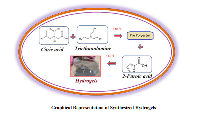
Hydrogels are polymeric networks capable of absorbing and retaining large amounts of water. These polymeric materials can swell and store a substantial volume of water within their structure while remaining insoluble in aqueous media. Due to their high-water content, hydrogels exhibit a high degree of flexibility, closely resembling natural tissues. Hydrogels absorb water due to the presence of hydrophilic groups within the polymer backbone [1]. Hydrogels are characterized by their ability to swell within the polymeric network. However, polymer–water interaction forces are the primary factor responsible for hydrogel swelling. Polymer–water interactions, electrostatic forces, and osmotic pressure play important roles in the swelling and expansion of the hydrogel polymeric network. Hydrogels can be categorized as non-ionic, ionic (anionic, cationic, and amphoteric), and hydrophilic hydrogels containing hydrophobic groups based on their swelling behavior. Non-ionic hydrogels, such as poly (N-vinyl pyrrolidone) and poly(ethylene oxide), swell in aqueous media due to polymer–water interactions. Hydrogels exhibit good transparency and can be easily modified. They are environmentally friendly and possess self-healing properties [2], enabling the timely delivery of drugs or nutrients [3]. Hydrogels have the ability to respond to changes in pH, temperature, metabolite concentration, and other environmental stimuli [4,5]. Chemical cross-linking enhances the mechanical strength of hydrogels [6]. This approach is highly effective for the formation of in situ hydrogel structures. Physical cross-linking techniques involve ionic interactions, as well as temperature-dependent and pH-dependent cross-linking mechanisms [7]. The wide range of applications includes tissue engineering and the design of biological tissues in both in vitro and in vivo systems [8,9], effective wound therapy [10], drug delivery [11], gene therapy [12], and the repair of blood vessels, skin, heart valves, cartilage, and tendons [13]. Additional applications include contact lens manufacturing [14], biosensors, and wastewater remediation techniques [15]. Zinc oxide (ZnO) nanoparticles have attracted considerable attention in biomedical applications due to their excellent antibacterial activity, biocompatibility, and stability. ZnO nanoparticles effectively inhibit the growth of a broad spectrum of microorganisms through the generation of reactive oxygen species and disruption of bacterial cell membranes. In addition to their antimicrobial properties, ZnO nanoparticles also promote wound healing by enhancing cell proliferation and tissue regeneration. Several metal and metal oxide nanoparticles, such as silver oxide (Ag₂O) [16], gold (Au), copper oxide (CuO) [17], titanium dioxide (TiO₂), magnesium oxide (MgO), and zinc oxide (ZnO) [18], have been extensively investigated for antibacterial and wound-healing applications due to their strong antimicrobial activity and ability to accelerate the healing process. Therefore, ZnO nanoparticles were incorporated into the CA4 matrix to prepare the CA4-ZnO nanocomposite, with the aim of enhancing its antibacterial activity and wound-healing potential. Biopolymeric three-dimensional hydrogel networks are formed from biocompatible components that can absorb and retain large quantities of water while maintaining their structural integrity. Owing to their biocompatibility, biodegradability, and tunable physicochemical properties, these hydrogels have gained significant attention in biomedical applications. In the present study, the hydrogel network was developed using citric acid, triethanolamine, and 2-furoic acid as functional building components. Citric acid acts as an effective cross-linking agent due to the presence of multiple carboxyl groups, which facilitate the formation of a stable polymeric network through esterification and hydrogen-bonding interactions. Triethanolamine contributes to the stabilization of the hydrogel structure through intermolecular interactions, while 2-furoic acid introduces additional functional groups that enhance the hydrogel’s physicochemical properties. To further enhance the functional performance of the hydrogel, zinc oxide (ZnO) nanoparticles were incorporated into the polymeric matrix to form a CA4-ZnO nanocomposite. The incorporation of ZnO nanoparticles into the hydrogel network enhances antimicrobial activity and promotes tissue regeneration, making the nanocomposite suitable for biomedical applications.
2.1. Methods 2.1.1. Stage I: Synthesis of Pre-Polymers Citric acid (CA) was used as the monomer. A quantity of 0.025 mol (4.803 g) of citric acid (CA) was dissolved in 5 mL of ethanol and then transferred into a round-bottom flask fitted with a mechanical stirrer until the monomer was completely dissolved in the ethanol. Triethanolamine (TEA) [0.025 mol (3.3006 g)], dissolved in 5 mL of ethanol, was added dropwise to the citric acid solution using a dropping funnel. The reaction mixture was stirred at 140 °C for 1 h. The formation of a sticky white gel indicated the formation of the citric acid–triethanolamine (CT) prepolymer). 2.1.2. Stage II: Synthesis of Bio-Polymeric Hydrogels 2-Furoic acid (FA) [0.025 mol (2.802 g)] was dissolved in 5 mL of ethanol, added to the CT prepolymer, and stirred continuously with a mechanical stirrer at 140 °C for 2 h. The formation of a glassy brown gel of citric acid–triethanolamine–2-furoic acid (CTF) confirmed the synthesis of the parent hydrogel. The resultant gel was immersed in pure ethanol for 24 h to remove unreacted monomers and subsequently dried in a vacuum oven at 35 °C for 24 h. A similar procedure was followed by altering the chemical composition of the monomers. Table 1 lists the experimental details of the synthesized biopolymeric hydrogels. Among the different hydrogel series, CA4 was selected for further investigation based on its superior swelling behavior compared with the other synthesized hydrogels. Formulation of CA-TEA-FA in distinct composition. 2.1.3. Incorporation of Zinc oxide (ZnO) Nanoparticles in CA4 Biopolymeric Hydrogels A total of 0.5 wt% zinc oxide (ZnO) nanoparticles with a particle size of less than 100 nm were incorporated into the CA4 biopolymeric hydrogel matrix and stirred continuously at 35 °C for 1 h, resulting in the formation of biopolymeric nanocomposite hydrogels [CA4-ZnO]. Similarly, 1.0 wt% and 2.0 wt% ZnO nanoparticles were separately incorporated into the CA4 biopolymeric hydrogels using the same procedure, as shown in Table 2. The ZnO concentrations (0.5, 1.0, and 2.0 wt%) were calculated based on the total weight of the synthesized polymer hydrogel matrix. The samples were named CA4Z1, CA4Z2, and CA4Z3 based on the composition of the CA4 hydrogel matrix and the ZnO nanoparticles. Here, CA4 represents the base hydrogel formulation (0.040 + 0.025 + 0.025 mol), Z indicates ZnO nanoparticles, whereas the numbers 1, 2, and 3 correspond to 0.5 wt%, 1.0 wt%, and 2.0 wt% ZnO nanoparticles, respectively. Formulations of CA–TEA–FA with different compositions. 2.2. Characterization 2.2.1. Fourier Transform Infrared (FTIR) Spectroscopic Analysis The molecular structure of the CA4-ZnO nanocomposite was characterized using a Shimadzu 8400S FTIR spectrophotometer. Samples were prepared using the standard KBr pellet method, and the spectra were recorded over the range of 4000–500cm−1. 2.2.2. UV–Vis Spectrophotometry An ultraviolet–visible double-beam (UV–Vis) spectrophotometer was used to characterize the CA4-ZnO nanocomposite. Samples of the compound were collected periodically to monitor the completion of the reaction. The sample spectra were recorded over a wavelength range of 190–1100 nm. 2.2.3. Scanning Electron Microscopy–Energy-Dispersive X-ray Spectroscopy (SEM -EDX) To evaluate the effects of polymer and cross-linker concentrations on the surface morphology, the CA4-ZnO hydrogel was characterized using scanning electron microscopy (SEM). SEM coupled with energy-dispersive X-ray spectroscopy (EDX) enabled rapid and accurate qualitative and quantitative analysis of the elemental composition, providing valuable insights into the hydrogel structure. 2.2.4. High-Resolution Transmission Electron Microscopy (HR-TEM) High-resolution transmission electron microscopy (HR-TEM), operated at 200–300 kV, was used to obtain high-resolution images and structural information at the nanoscale. 2.2.5. Thermogravimetric Analysis (TGA) The thermal properties of the polymeric hydrogels were investigated using simultaneous thermogravimetric analysis (TGA) with an SDT Q600 (TA Instruments). The TGA curves were recorded over a temperature range from room temperature to 500 °C. 2.2.6. Swelling Behavior A 0.200 g sample of dried hydrogel was immersed in buffer solutions with pH values ranging from 2.0 to 11.0 at room temperature. At regular time intervals, the swollen hydrogels were carefully removed from the swelling medium, gently blotted with filter paper to remove excess surface water, weighed, and then returned to the original swelling medium. This procedure ensured the accurate determination of the water absorption capacity. Finally, excess surface water was removed from the hydrogels with filter paper. The swelling percentage was calculated using Equation (1): where, WI is the initial weight of the dried hydrogel and WSq is the weight of the swollen sample at equilibrium. 2.3. Biological Applications 2.3.1. Antibacterial Studies The antibacterial activity was evaluated using the conventional agar well diffusion method. Each bacterial isolate was cultured in brain heart infusion (BHI) broth and diluted to approximately 10⁵ colony-forming units (CFU)/mL. The bacterial suspensions were flood-inoculated onto the surface of Mueller–Hinton agar and allowed to dry. Sterile cork borers were used to create wells with a diameter of 5 mm in the agar medium. Subsequently, 30 µL of the sample solution (50 µg compound dissolved in 500 µL DMSO) was placed in each well. The plates were incubated at 37 °C for 18 h. Antimicrobial activity was evaluated by measuring the diameter of the inhibition zone. Dimethyl sulfoxide (DMSO) was used as the solvent control, while ciprofloxacin was used as the standard antibacterial agent. The tests were performed in triplicate. The hydrogels were tested against Staphylococcus aureus, Bacillus subtilis, Klebsiella pneumoniae, and Escherichia coli. 2.3.2. Antifungal Studies The antifungal activity was evaluated against C. albicans and A. niger. Clotrimazole (20 mg/well) was used as the standard. The diameter of the inhibition zone (mm) was used as an indicator of activity against the test pathogens. 2.3.3. Cytotoxic Studies The mouse fibroblast cell line (L929) was seeded in a 96-well plate at a concentration of 1 × 10⁴ cells/well in Dulbecco’s Modified Eagle Medium (DMEM) supplemented with 10% fetal bovine serum and 1× antibiotic–antimycotic solution. The cells were incubated in a CO₂ incubator maintained at 37 °C with 5% CO₂. After washing with 200 μL of 1× phosphate-buffered saline (PBS), the cells were cultured for 24 h in serum-free medium containing different concentrations of the test sample, with 25% DMSO used as the positive control. At the end of the treatment period, the culture medium was aspirated from the cells. MTT solution [0.5 mg/mL of (3-(4,5-dimethylthiazol-2-yl)-2,5-diphenyltetrazolium bromide)] prepared in 1X PBS was added, and the cells were incubated for 4 h at 37 °C in a CO2 incubator. After the incubation period, the medium containing MTT was discarded, and the cells were rinsed with 200 μL of PBS. The formed crystals were dissolved in 100 μL of DMSO and mixed thoroughly. The formation of purple-blue formazan dye was quantified by measuring the absorbance at 570 nm using a microplate reader. 2.3.4. Antioxidant Studies Blois’s (1958) method was used to measure the sample’s DPPH (2,2-diphenyl-1-picrylhydrazyl) radical-scavenging activity. A 0.5 mL aliquot of the sample solution in methanol was mixed with 2.5 mL of a 0.5 mM methanolic DPPH solution. The reaction mixture was shaken well and kept in the dark at room temperature for 30 min. The absorbance was measured at 517 nm using a UV spectrophotometer. Ascorbic acid was used as the positive control. The percentage of DPPH free radical scavenging activity was determined using the standard formula. 2.3.5. Computational Studies A preliminary computational study was conducted to investigate the antibacterial activity and protein-binding properties of CA4 hydrogels and ZnO nanoparticle-incorporated hydrogels. Therefore, the CA4-ZnO nanocomposite hydrogels were subjected to molecular docking studies.Table 1: S. No
Samples
Monomer
Composition (mole)Description of Hydrogels
CA
TEA
FA
1
CTF
0.025
0.025
0.025
Transparent in nature-
Brown glassy gel-
Insoluble in water2
FA1
0.025
0.025
0.01
3
FA2
0.025
0.025
0.02
4
FA3
0.025
0.025
0.03
5
FA4
0.025
0.025
0.04
6
TEA1
0.025
0.01
0.025
7
TEA2
0.025
0.02
0.025
8
TEA3
0.025
0.03
0.025
9
TEA4
0.025
0.04
0.025
10
CA1
0.01
0.025
0.025
11
CA2
0.02
0.025
0.025
12
CA3
0.03
0.025
0.025
13
CA4
0.04
0.025
0.025
Table 2: S. No
Sample
CA4 Hydrogel Composition (moles)
ZnO
Nanocomposite (wt %)Description of Hydrogels
1
CA4Z1
0.040 + 0.025 + 0.025
0.5
Transparent in nature; brown, glassy gel; insoluble in water
2
CA4Z2
0.040 + 0.025 + 0.025
1.0
3
CA4Z3
0.040 + 0.025 + 0.025
2.0
(1)
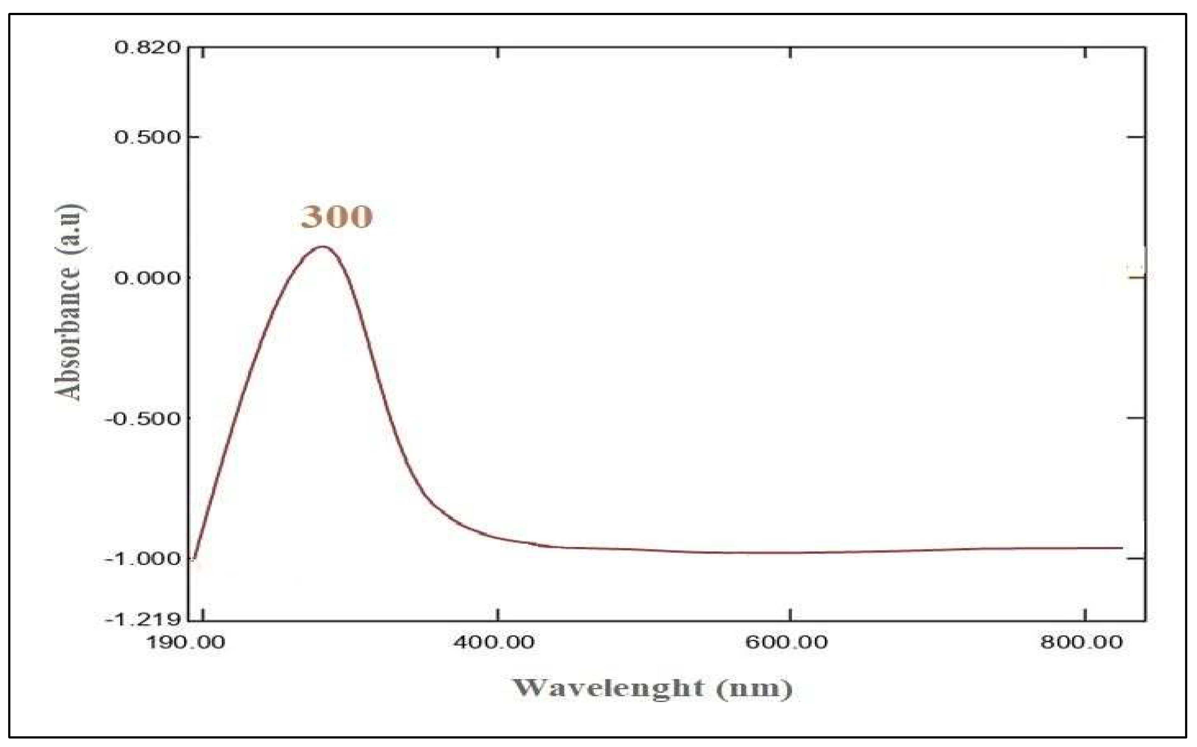
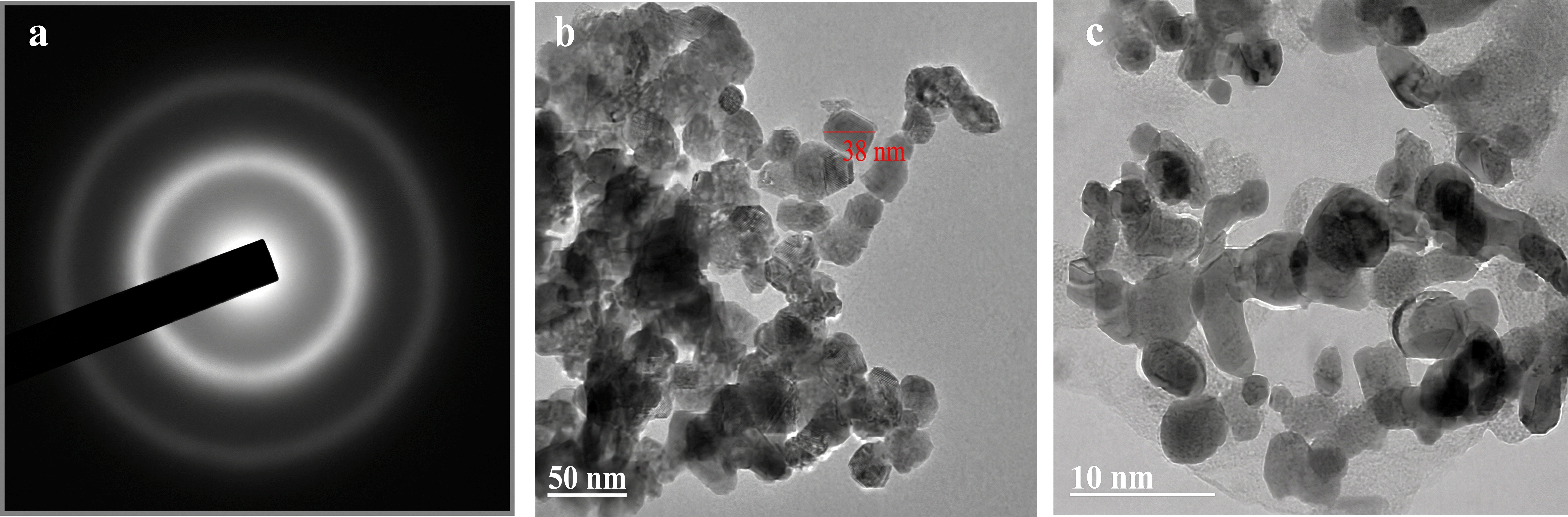
3.1. Spectral Characterization 3.1.1. FTIR Spectroscopy of CA4-ZnO Nanocomposite Hydrogels A stretching frequency observed at 3360 cm−1 corresponds to the hydrogen-bonded O-H stretching vibration [19,20]. The decrease in the stretching frequency from 3414 cm−1 to 3360 cm−1 may be attributed to the incorporation of ZnO nanoparticles into the polymer network. The peak at 2982 cm−1 represents the C-H stretching vibration. A distinct sharp peak at 1732 cm−1 is ascribed to C=O stretching [21]. New absorption peaks at 1610 cm−1 and 1474 cm−1 were attributed to COO− stretching vibrations. The prominent spectral peak at 1294 cm−1 is due to the presence of C-N stretching vibrations found in triethanolamine. The C-O stretching vibration was observed at 1176 cm−1 in the polyester network. The peaks at 1073 cm−1 and 1041 cm−1 were attributed to C-O-C stretching vibrations. The spectral peak appeared at 670 cm−1, confirming the incorporation of zinc oxide nanoparticles into the main chain of hydrogels, as shown in Figure 1 [22]. 3.1.2. UV Spectroscopy of [CA4-ZnO] Nanocomposite Hydrogels UV–Vis spectroscopy is a key analytical technique used to characterize citric acid-, 2-furoic acid-, and triethanolamine-based hydrogels incorporated with zinc oxide (ZnO) nanoparticles. This method helps confirm the incorporation of ZnO nanoparticles into the hydrogel matrix. Figure 2 shows the UV–Vis spectral findings of CA4-ZnO nanocomposite hydrogels. ZnO nanoparticles absorb UV–Vis light over the wavelength range of 250–800 nm [23]. The UV-Visible spectrum of the CA4-ZnO nanocomposite revealed a broad absorption peak at 300 nm, corresponding to an absorbance value of 0.1. The absorbance of a nanocomposite hydrogel is caused by n→π* transitions. The λmax primarily corresponds to chromophores containing carbonyl groups. This transition involves the promotion of an electron from a nonbonding (n) orbital, specifically a lone pair on a heteroatom such as oxygen, to an antibonding (π*) orbital. 3.1.3. SEM and EDX Analysis of CA4-ZnO Nanocomposite Hydrogels The surface morphology of the synthesized CA4-ZnO nanocomposite hydrogel was examined using the CA4Z3 sample by scanning electron microscopy (SEM). The SEM image (Figure 3) reveals a heterogeneous, nonporous surface morphology with a rough structure attributed to the hydrogel matrix containing ZnO nanoparticles, which are irregularly distributed throughout the network. The interaction between the hydrogel polymer chains and ZnO nanoparticles enhances the swelling ability and functional properties of the hydrogel. To validate the synthesis of the CA4-ZnO nanocomposite hydrogel CA4Z3, EDX analysis was performed, and the resulting peaks are shown in Figure 4. The EDX analysis confirmed the presence of ZnO nanoparticles in the synthesized nanocomposite. The elemental composition of the nanocomposite, expressed in weight percentage, revealed the presence of carbon (C), oxygen (O), nitrogen (N), and zinc (Zn) at 56.16%, 30.69%, 3.29%, and 7.51%, respectively. The plot of CA4-ZnO nanocomposite hydrogels shows the presence of carbon, nitrogen, oxygen, and zinc (Table 3). Weight percentages of elements in the CA4Z3 nanocomposite hydrogel. 3.1.4. HR-TEM Analysis of CA4-ZnO Nanocomposite Hydrogels The size and structure of nanoparticles within the hydrogel network were examined using transmission electron microscopy (TEM). The TEM images showed that the spherical ZnO nanoparticles were uniformly distributed within the CA4-ZnO hydrogel matrix. The average particle size of the CA4-ZnO nanocomposite was approximately 38 nm (Figure 5). The well-defined spherical morphology of the ZnO nanoparticles indicates strong interactions between the hydrogel matrix and ZnO nanoparticles. Furthermore, the presence of lower concentrations of spherical ZnO nanoparticles enhanced the biological activity, as evidenced by the antibacterial studies [24]. 3.1.5. TGA of [CA4-ZnO] Nanocomposite Hydrogels Thermogravimetric analysis (TGA) was performed to investigate the thermal stability of the CA4-ZnO nanocomposite hydrogel, as shown in Figure 6. The TGA curve shows three stages of degradation. The initial stage of degradation occurred between 50 °C and 100 °C, resulting in a 9% weight loss due to the removal of residual water. The second stage of degradation occurred between 150 °C and 250 °C, resulting in a 23% weight loss due to the decomposition of 2-furoic acid substituents from the hydrogel matrix (Figure 6). The third stage of degradation was observed at 300 °C, resulting in a 16% weight loss due to the decomposition of the polymer network containing embedded ZnO nanoparticles [25]. Overall, the TGA study revealed that the ZnO nanocomposite hydrogel exhibited considerable thermal stability, with several degradation stages involving water loss and polymer decomposition. In general, ZnO nanoparticles exhibit high thermal stability under normal conditions. However, their stability can be affected by factors such as impurities, interactions with other materials, and exposure to high temperatures, which may lead to structural degradation. In the present study, the interaction between the biopolymeric hydrogel matrix and ZnO nanoparticles resulted in reduced thermal stability, likely due to polymeric filler interactions [26]. 3.1.6. Swelling Equilibrium (%) of CA4-ZnO Nanocomposite Hydrogels ZnO nanoparticles at concentrations of 0.5%, 1.0%, and 2.0%, designated as CA4Z1, CA4Z2, and CA4Z3, respectively, were incorporated into CA4 hydrogels. The swelling behavior of the CA4-ZnO nanocomposite hydrogels at various pH values (2.0, 4.0, 7.0, 9.0, and 11.0) was evaluated, and the results are presented in Table 4. The hydrogels exhibited pH-dependent swelling, with maximal swelling observed under neutral to slightly alkaline conditions (pH 7–9), owing to the ionization of functional groups such as -COOH and -OH in the polymeric matrix, which increases electrostatic repulsion and favors higher water uptake. CA4Z3 had the largest swelling percentage (127% at pH 7), followed by CA4Z2 (121%) and CA4Z1 (117%), as shown in Figure 7. Increasing the concentration of ZnO nanoparticles appears to enhance the swelling capacity, which may be attributed to their interactions with the hydrophilic functional groups within the hydrogel network. At acidic pH (2–4), the swelling ratios decreased due to protonation of functional groups, which promoted hydrogen bonding and network contraction. At a higher alkaline pH of 11, swelling was partially reduced due to charge shielding and partial degradation of the hydrogel network [27]. Overall, the swelling investigation demonstrates that the incorporation of ZnO nanoparticles into the hydrogel is advantageous for applications in drug delivery, wound dressing, and biomedical systems requiring controlled release under physiological pH conditions [28]. The biological activities in the present study were evaluated using CA4Z3 nanocomposite hydrogels due to their favorable swelling behavior. Swelling equilibrium (%) of CA4-ZnO at different pH values. 3.2. Biological Activity of [CA4-ZnO] Nanocomposite Hydrogels 3.2.1. Antibacterial Activity CA4Z3 outperformed all other [CA4-ZnO] hydrogel compositions in terms of antibacterial performance, as shown in Figure 8. The antibacterial activity was evaluated against Gram-positive bacterial strains (S. aureus and B. subtilis) and Gram-negative bacterial strains (E. coli and K. pneumoniae), using ciprofloxacin as the standard reference drug. The standard drug ciprofloxacin produced inhibition zones of 25 mm against S. aureus, 18 mm against B. subtilis, 20 mm against K. pneumoniae, and 30 mm against E. coli. The CA4Z3 hydrogels exhibited inhibition zones of 0 mm against E. coli, 8 mm against S. aureus, 10 mm against B. subtilis, and 6 mm against K. pneumoniae (Figure 8). These findings indicate that the hydrogel exhibited moderate inhibitory activity against S. aureus, B. subtilis, and K. pneumoniae, whereas E. coli showed no antibacterial activity, which may be attributed to variations in cell membrane permeability toward the antibacterial agent [29]. The obtained results demonstrate that CA4Z3 hydrogels exhibited moderate inhibitory effects, indicating their potential for biological applications. 3.2.2. Antifungal Activity The hydrogels were evaluated for antifungal activity against fungal strains, including Candida albicans and Aspergillus niger, using the agar well diffusion method. The standard antifungal drug clotrimazole exhibited inhibition zones of 16 mm against Candida albicans and 26 mm against Aspergillus niger. In comparison, the CA4Z3 hydrogel at a concentration of 300 mg/well produced a 4 mm inhibition zone against Candida albicans, while no inhibition (0 mm) was observed against Aspergillus niger (Figure 9). These findings revealed that the antifungal activity of the hydrogel was significantly lower than that of the standard drug. The limited inhibition may be attributed to the robust chitinous cell walls and melanin pigments present in these fungi, which enhance resistance to oxidative stress and reduce the susceptibility of the cells to ZnO nanoparticle attack [30]. Nevertheless, the incorporation of ZnO nanoparticles into polymeric hydrogel matrices may provide a synergistic antifungal platform with potential applications in biomedical and agricultural fields. 3.2.3. Cytotoxicity Activity Several studies have investigated the cytotoxicity of ZnO nanoparticles using the MTT assay, demonstrating their effects on a wide range of animal cell types [31]. Figure 10 shows the cell viability% of CA4Z3 nanocomposite hydrogels. The concentrations of 25, 50, 100, 250, and 500 µg/mL yielded percentages of 95, 90, 90, 85, and 82%, respectively. CA4Z3 was classified as non-toxic according to the criteria defined by ISO 10993-5 [32]. The average cell viability was greater than 80% at all concentrations. As the result, among the CA4-ZnO nanocomposite hydrogels, CA4Z3 exhibited a higher percentage of cell viability, particularly at lower concentrations (25, 50, and 100 µg/mL), indicating its suitability and safety for biomedical applications (Figure 11). 3.2.4. Antioxidant Activity The antioxidant activity of CA4Z3 nanocomposite hydrogels was evaluated using the DPPH radical-scavenging assay, with ascorbic acid as the reference standard. The inhibition rates ranged from 32% to 94% at different concentrations (25, 50, 100, 250, and 500 µg/mL), with corresponding values of 32%, 42%, 67%, 90%, and 94%, respectively. CA4Z3 hydrogels exhibited antioxidant activity of 42%, 53%, 61%, 68%, and 79% at concentrations of 25, 50, 100, 250, and 500 µg/mL, respectively, as shown in Figure 12. At lower concentrations (25 and 50 μg/mL), CA4Z3 exhibited moderate inhibition, whereas ascorbic acid showed comparatively lower inhibition. At medium concentrations (100–250 µg/mL), the antioxidant activity of CA4Z3 gradually increased, followed by a further increase in activity at higher concentrations. At the maximum concentration (500 µg/mL), CA4Z3 exhibited good inhibition compared with ascorbic acid. These results suggest that the ZnO nanocomposite hydrogel possesses significant antioxidant properties, which may be attributed to the interaction of ZnO nanoparticles with free radicals. This may promote electron donation and hydrogen transfer to DPPH radicals through the hydrophilic functional groups (-OH, -NH, and -COOH) present in the hydrogel matrix. The interaction between ZnO nanoparticles and the polymeric network facilitates enhanced radical-scavenging efficacy. The IC50 of CA4Z3 was 149 µg/mL for the DPPH method. The scavenging activity and IC50 value exhibit an inverse relationship [33]. Similar findings have also been reported by Patel et al. (2011) [34]. 3.3. Computational Studies of CA4-ZnO Nanocomposite Hydrogels 3.3.1. Density Functional Theory (DFT) Since the CA4-ZnO nanocomposite hydrogels exhibited biological activity, DFT and molecular docking studies were conducted to explore their mechanism of action. Initially, an intrinsic reaction coordinate (IRC) analysis was performed using the three basic monomers, namely citric acid, triethanolamine, and 2-furoic acid, which were utilized in the synthesis of the hydrogels. The IRC study revealed the structure selected for optimization at the DFT level. The monomers were further optimized at the 6-31G(d) level using the B3LYP (Becke, 3-parameter, Lee–Yang–Parr) functional, and the FMOs (Frontier Molecular Orbitals) were visualized to evaluate the molecular orbital aspects of reactivity. After geometry optimization, the molecular electrostatic potential (MESP) was generated to identify probable reactive regions of the molecule. The optimized molecules were subsequently used for docking studies. The DFT-derived values for the CA4-ZnO nanocomposite hydrogels are presented in Table 5. The values generated from DFT calculations of CA4-ZnO nanocomposite hydrogels. The FMO analysis revealed that, although the three monomers combine to form the hydrogel network, the IRC results indicated that the FMOs were unevenly distributed across the individual monomers. After optimization, it was observed that the HOMO (Highest Occupied Molecular Orbital) was primarily localized on the triethanolamine moiety of the hydrogel, whereas the LUMO (Lowest Unoccupied Molecular Orbital) was distributed slightly over the citric acid moiety, in close proximity to the ZnO nanoparticles within the hydrogel network (Figure 13). The 2-furoic acid moiety exhibited no lobes over its structure, indicating the absence of significant reactive regions within this segment. The denser lobes observed near the ZnO nanoparticle region suggest the presence of prominent reactive regions around its center. The MESP illustrates the distribution of electrostatic potential across the molecular surface, including the presence of positive potential regions, as shown in Figure 14 and Figure 15. These findings suggest that the corresponding moiety contributes to the desired biological activity through electronic effects arising from the coexistence of negative and positive potential regions. A higher-potential region was observed near the ZnO nanoparticle region, and the biological activity was enhanced through the incorporation of ZnO nanoparticles into the hydrogel matrix. 3.3.2. Molecular Docking (PDB ID: 1N67 and 4DXD) In view of their biomedical properties, the nanocomposite hydrogels were subjected to docking studies against important receptors associated with antibacterial activity and wound healing, including the FtsZ (filamenting temperature-sensitive mutant Z) protein complex [35] and the Staphylococcus aureus protein target Clumping Factor A [36]. Infections caused by drug-resistant strains of Klebsiella pneumoniae represent a major challenge to the efficacy of conventional antibiotics. Therefore, there is an increasing need to investigate alternative antimicrobial therapies, particularly hydrogels synthesized from simple monomeric compounds [37]. Considering the biological importance of hydrogel derivatives, the present study aimed to synthesize, characterize, and investigate the biological potential of the hydrogel using a simple, convenient, and eco-friendly synthetic methodology. The molecular docking study was conducted to evaluate the interactions between CA4-ZnO nanocomposite hydrogels and the FtsZ protein complex (PDB ID: 4DXD), given their strong antibacterial properties. Additionally, CA4-ZnO nanocomposite hydrogels were evaluated against the Staphylococcus aureus protein Clumping Factor A (PDB ID: 1N67), which demonstrated significant antibacterial activity. Both nanocomposite hydrogels exhibited favorable docking scores. However, the CA4-ZnO nanocomposite hydrogel demonstrated stronger and more favorable binding interactions with the Clumping Factor A protein, as shown in Table 6. The CA4-ZnO nanocomposite hydrogel also demonstrated superior ligand efficiency across all evaluated parameters, including standard ligand efficiency (−0.188), surface area-adjusted ligand efficiency (−0.633), and log-normalized ligand efficiency (−1.543). Docking properties of CA4-ZnO nanocomposite hydrogels against Clumping Factor A. In the CA4-ZnO–Clumping Factor A complex, the ligand is tightly anchored via hydrogen bonds with polar residues (ASN267, SER268, and GLN235) and stabilized by hydrophobic contacts (VAL270, VAL323, ALA269, and ILE232) and potential electrostatic interactions with ASP273. Water-mediated interactions and minimal solvent exposure further enhance binding stability. These findings suggest that CA4-ZnO nanocomposite hydrogels may serve as effective modulators of S. aureus proteins. 3.3.3. ADMET (Absorption, Distribution, Metabolism, Excretion, and Toxicity) The water solubility of a compound (LogS) represents the solubility of the compound at 25 °C. Drugs with higher water solubility are absorbed more efficiently than lipid-soluble compounds, and the obtained value of −2.99 indicates favorable aqueous solubility. Similarly, intestinal absorption predicts the percentage of an orally administered drug that is absorbed. The low absorption value observed in this study suggests that the compound may be more effectively administered through alternative routes rather than oral delivery. The blood-brain barrier (BBB) indicates the ability of a drug to penetrate the brain and represents an important factor in minimizing side effects and toxicity while enhancing therapeutic efficacy. The evaluated nanocomposite hydrogels were predicted to cross the BBB, as presented in Table 7. The obtained value suggests favorable BBB permeability for the investigated drug delivery system. ADMET properties of CA4-ZnO nanocomposite hydrogels.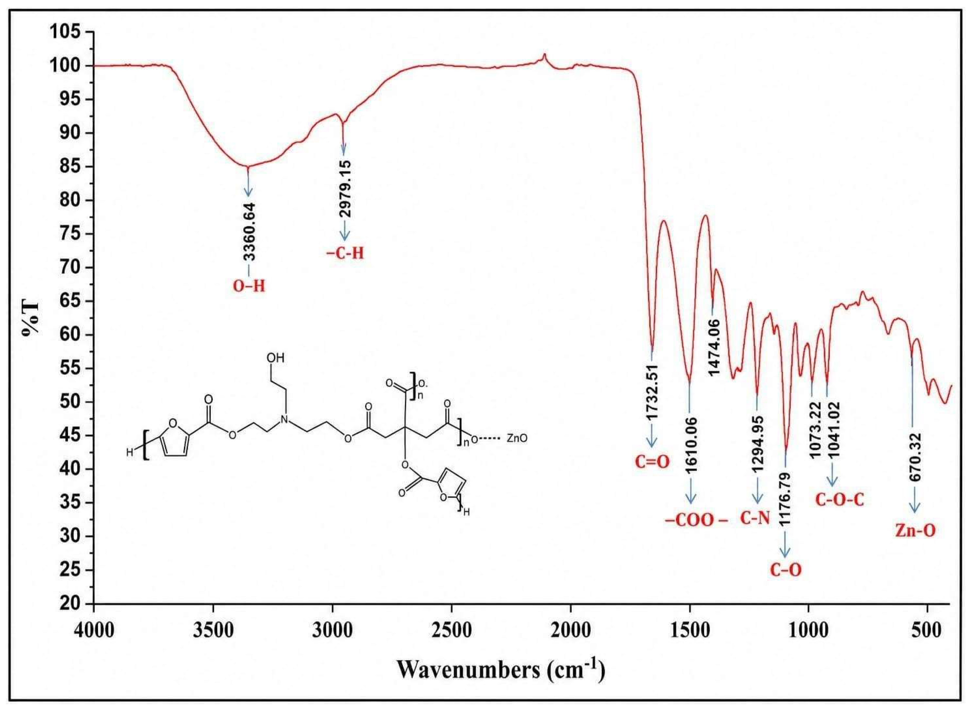
![Figure 3: <p>SEM analysis of [CA4-ZnO] nanocomposite hydrogels of CA<sub>4</sub>Z<sub>3</sub>.</p>](/uploads/source/articles/ecological-sustainable-materials-connect/2026/volume1/20260003/image003.png)
![Figure 4: <p>EDX analysis of [CA<sub>4</sub>-ZnO] nanocomposite hydrogels of CA<sub>4</sub>Z<sub>3</sub>.</p>](/uploads/source/articles/ecological-sustainable-materials-connect/2026/volume1/20260003/image004.png) Table 3:
Table 3: S. No
Elements
Atomic number
Weight percentage
1
Carbon
6
56.16
2
Oxygen
8
30.69
3
Nitrogen
7
3.29
4
Zinc
30
7.51
![Figure 6: <p>TGA of [CA<sub>4</sub>-ZnO] nanocomposite hydrogels.</p>](/uploads/source/articles/ecological-sustainable-materials-connect/2026/volume1/20260003/image006.png) Table 4:
Table 4: S. No
Sample
CA4
Synthesized HydrogelZnO Nano
Particles
(wt%)Swelling Equilibrium (%)
pH
(in Grams)
2
4
7
9.0
11
1
CA4Z1
0.200
0. 5
100
107
118
104
100
2
CA4Z2
0.200
1.0
108
113
120
115
107
3
CA4Z3
0.200
2.0
112
117
127
121
117
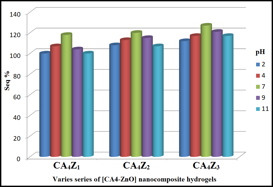
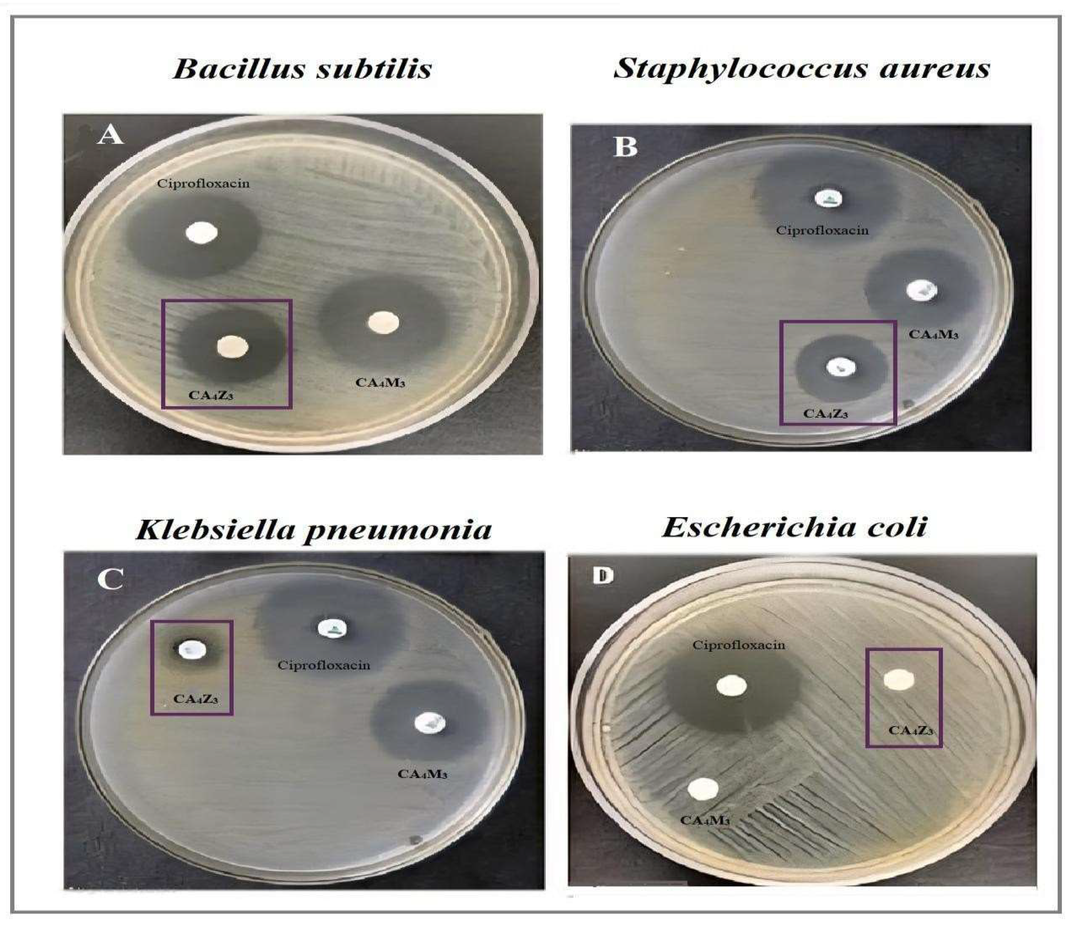
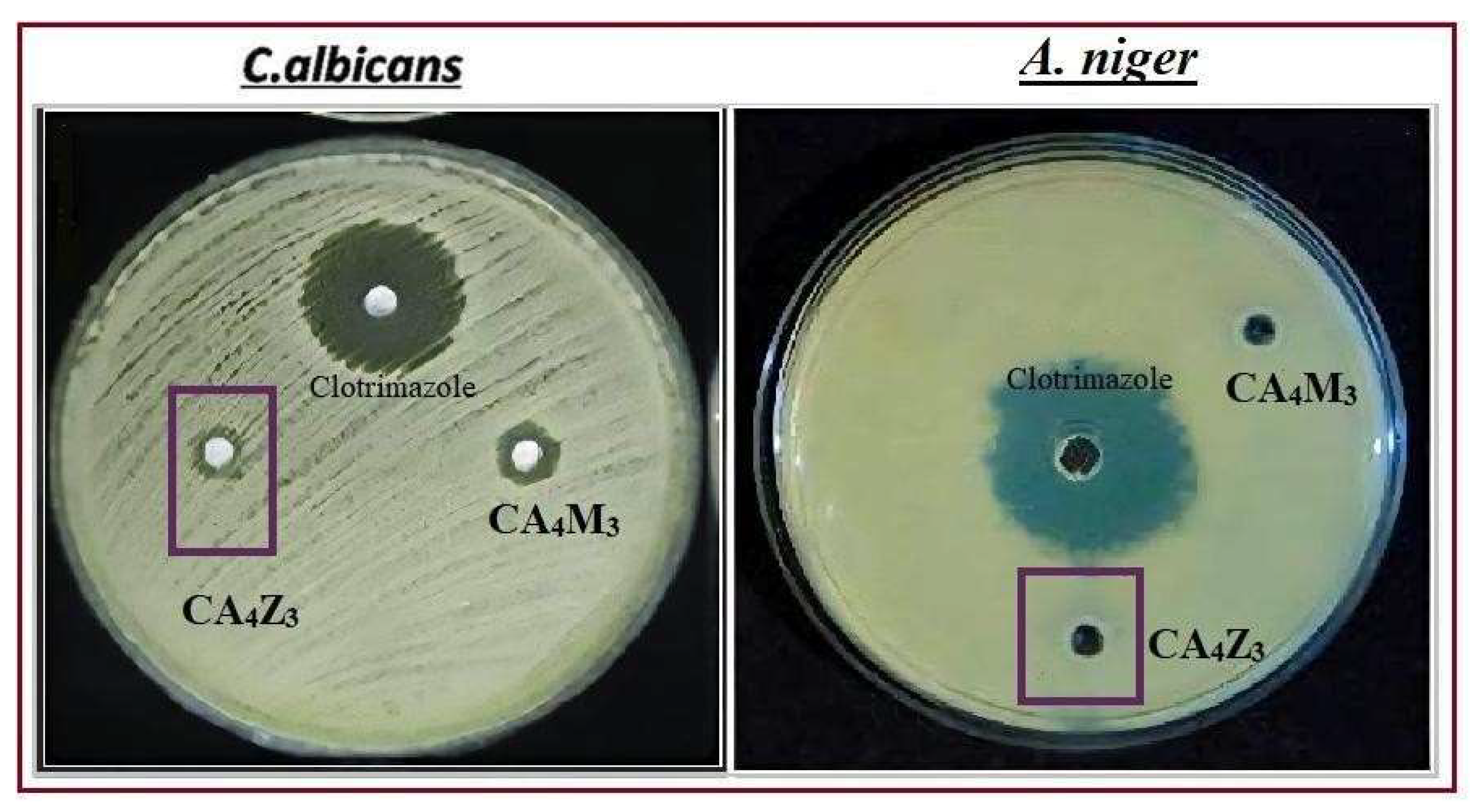
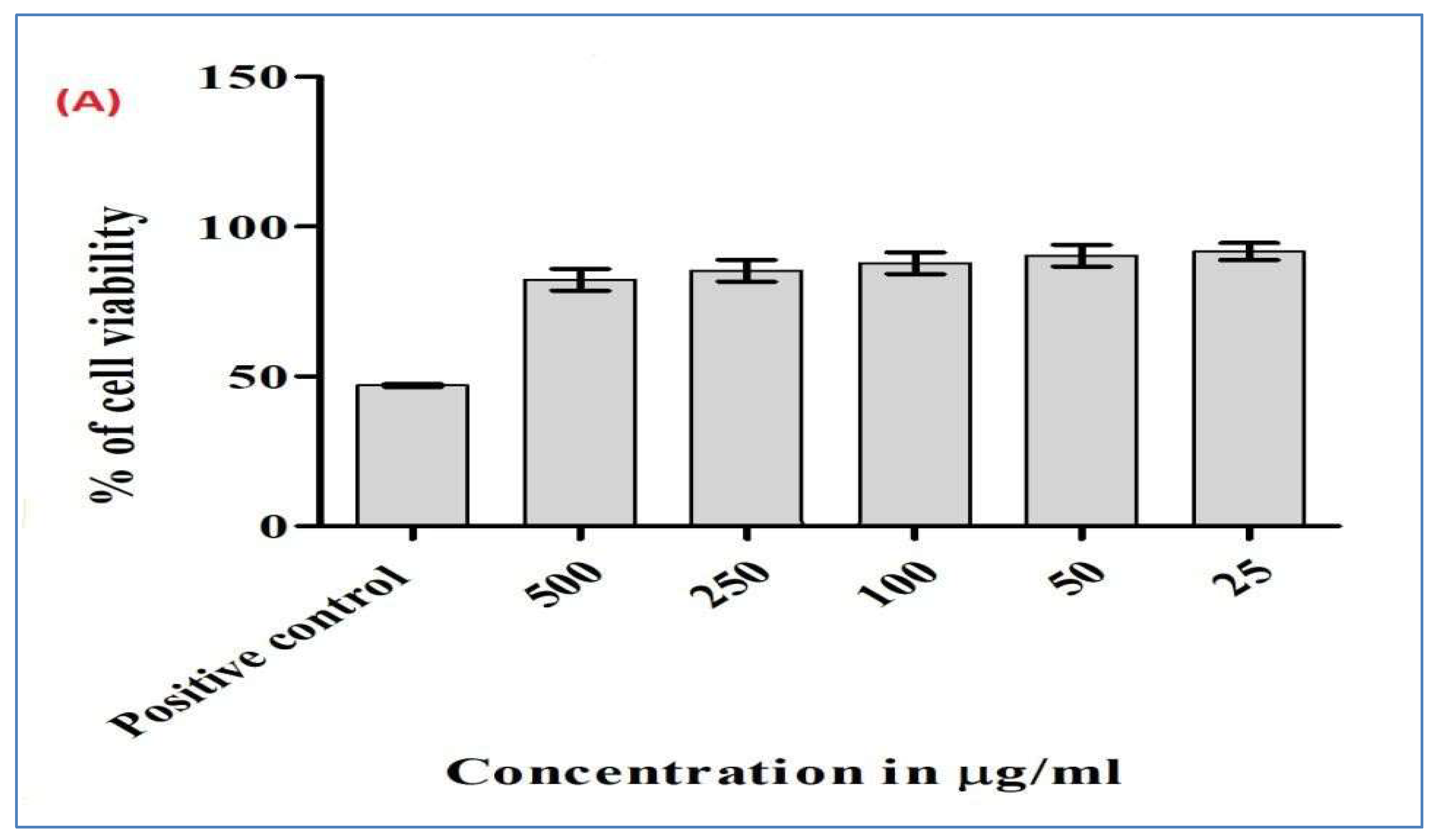
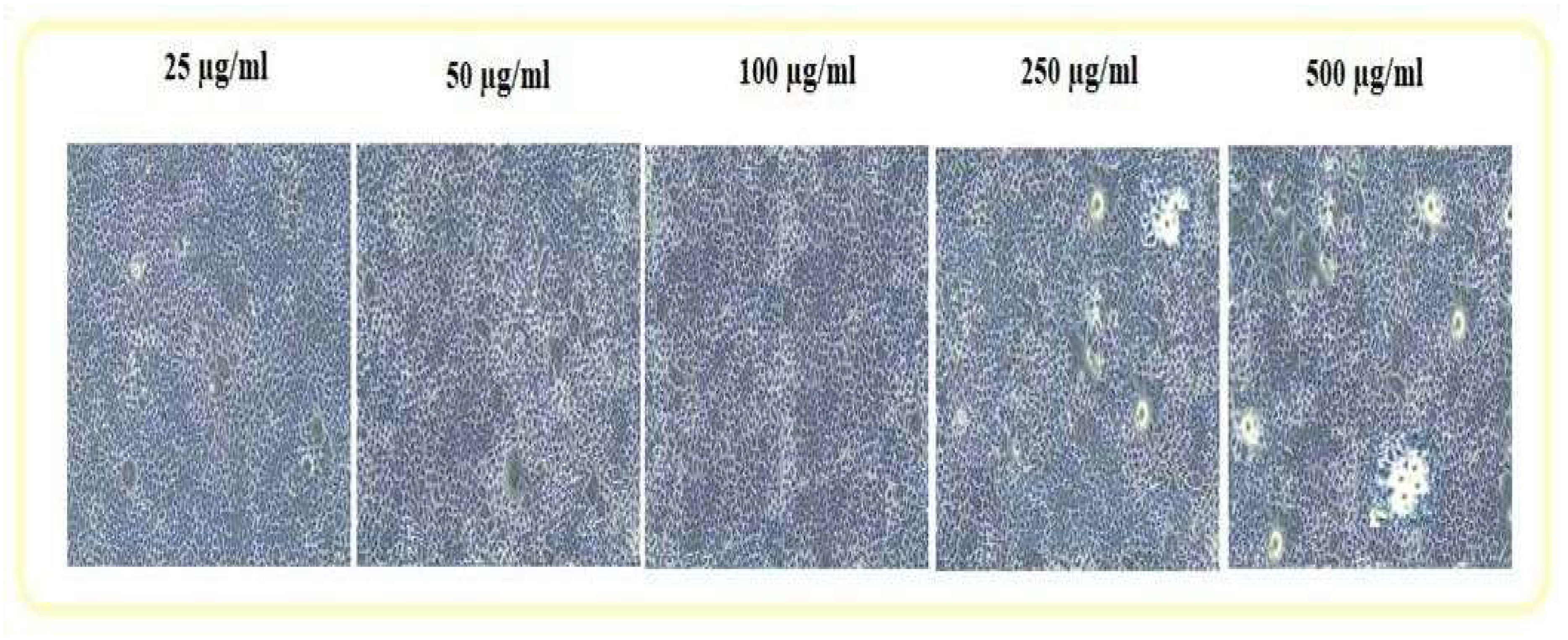
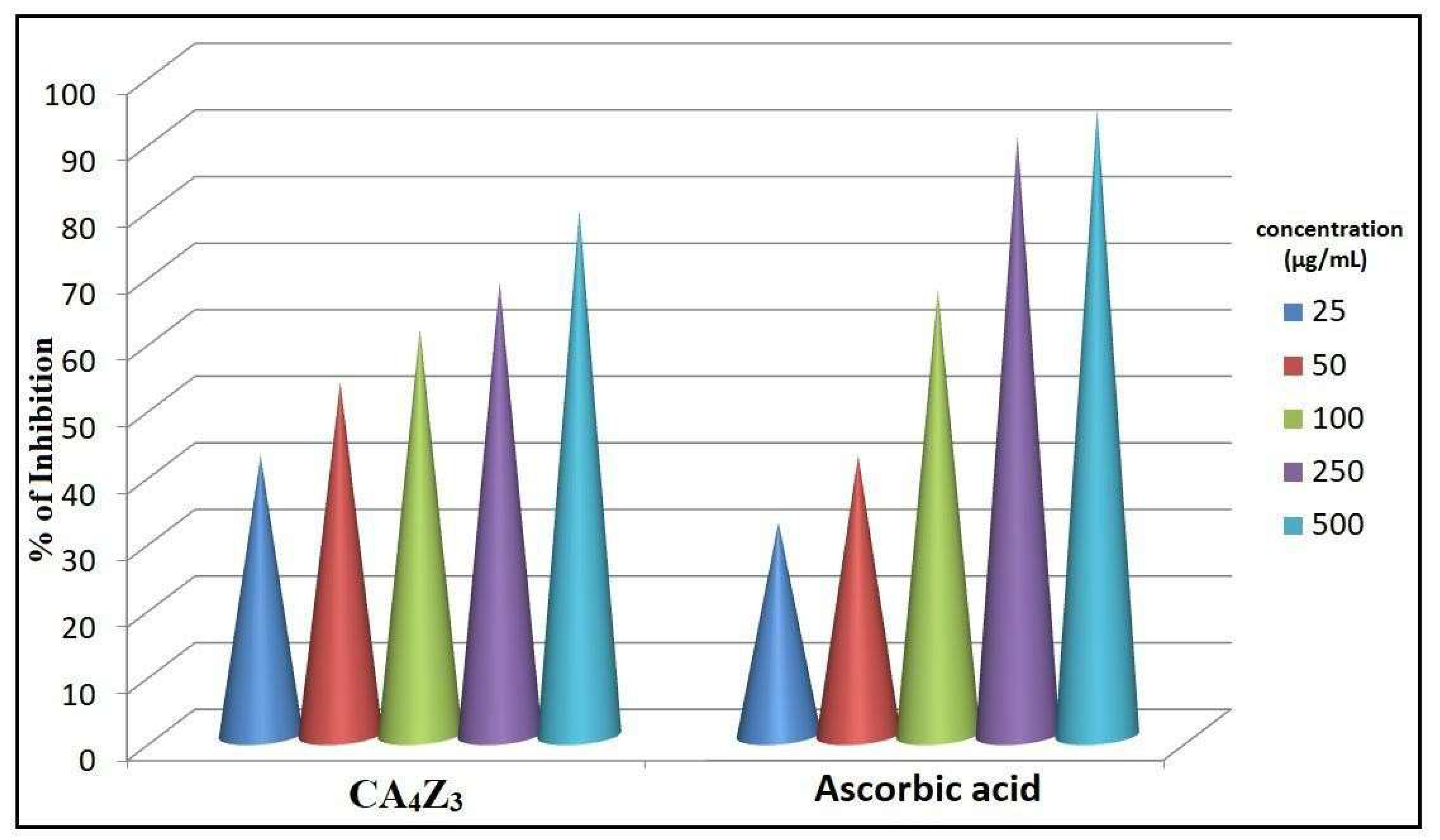 Table 5:
 Description
HOMO
eVLUMO
eVBand
Gap eVEnergy
HDipole D
Symmetry
CA4-ZnO
−6.171
−2.086
4.085
−3740.41
7.8291
C1
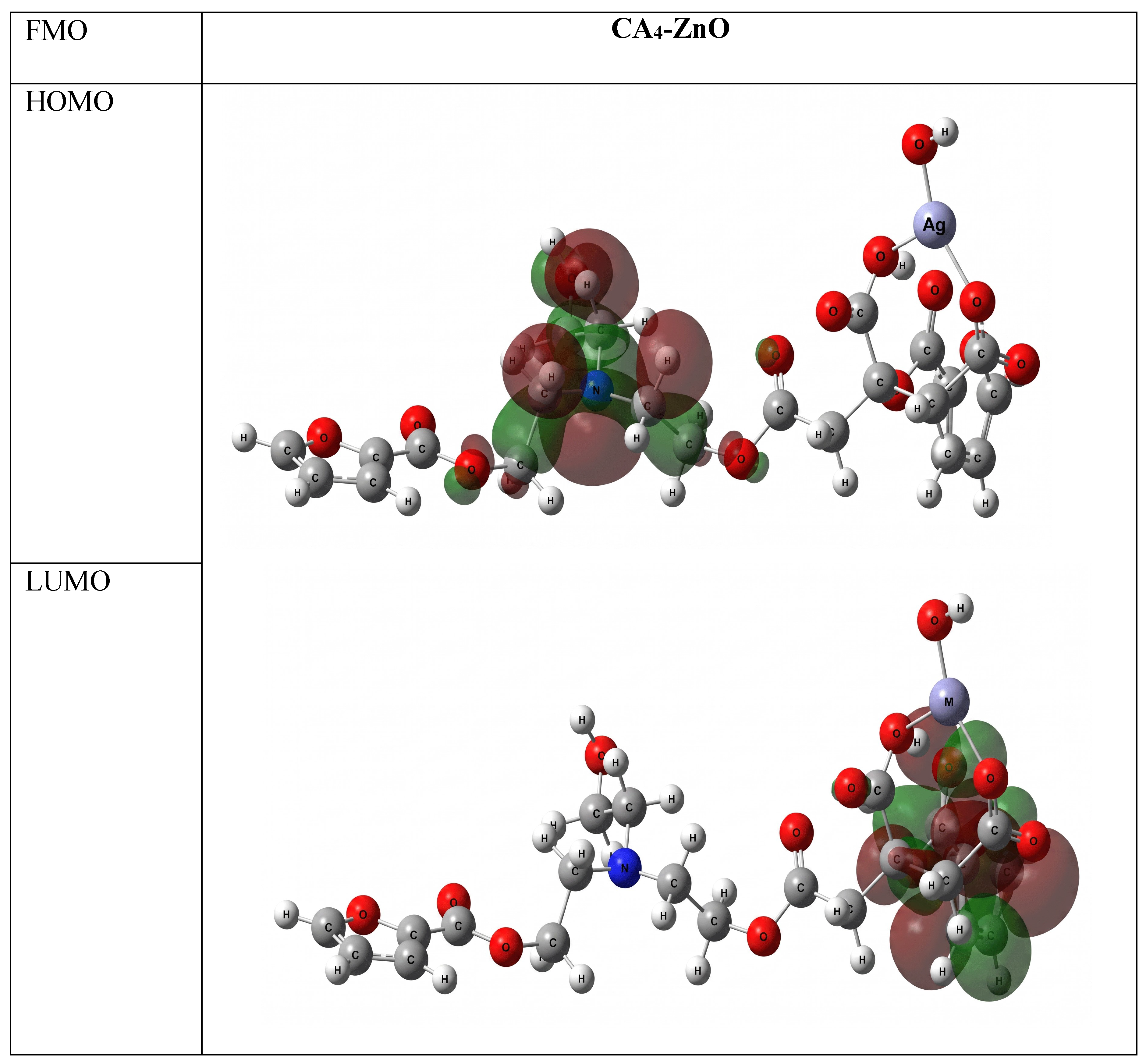
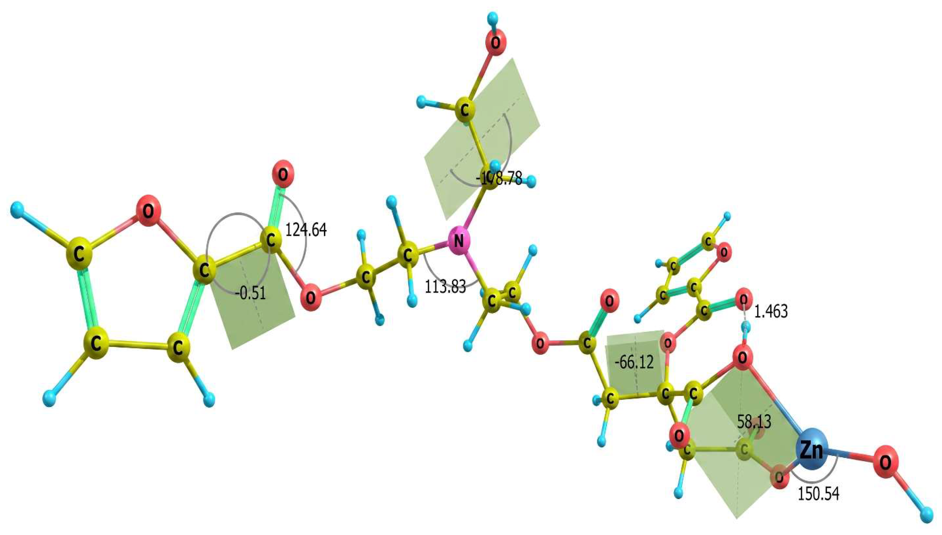
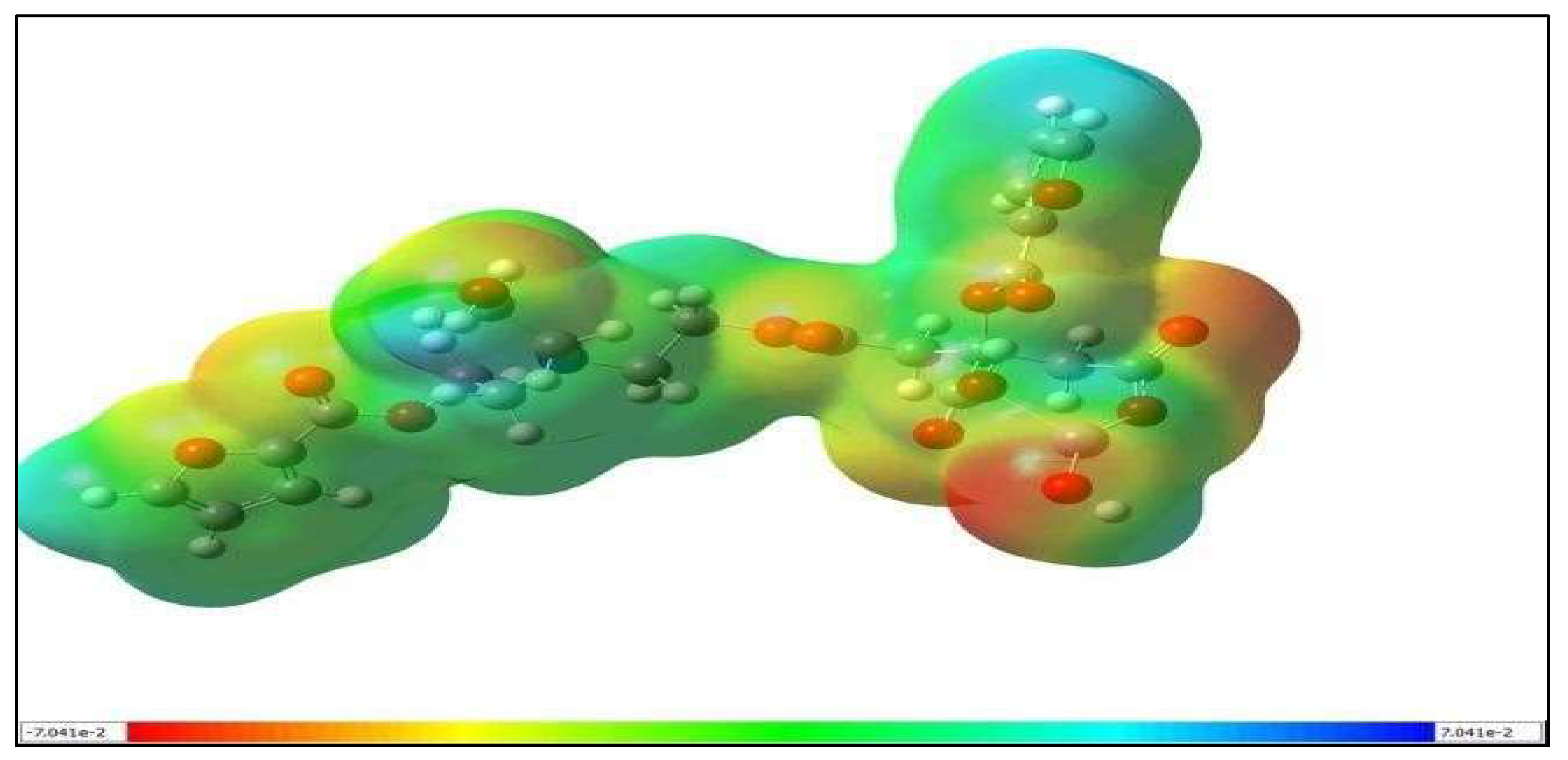 Table 6:
 Property
Clumping Factor a From Staphylococcus Aureus (1N67) CA4-ZnO
Docking Score
−7.154
Glide Ligand Efficiency
−0.188
Glide Ligand Efficiency SA
−0.633
Glide Ligand Efficiency LN
−1.543
Glide G Score
−7.154
Glide Evdw
−21.263
Glide Ecoul
−21.754
Glide Emodel
−42.555
Glide Energy
−43.017
XP GScore
−7.154
XP HBond
−1.860
Table 7: Properties
CA4-ZnO
Molecular Weight
592.82
Water Solub
−2.99
Caco2 perm
−0.23
Intest Abs
32.4
Skin Perm
−2.73
Glycopro substrate
Yes
BBB Perm
−2.43
CNS Perm
−3.85
CYP1A inhibitor
No
Log P
0.624
Surface area
223.12
In the current research, pH-responsive CA4 hydrogels with varying nano-ZnO concentrations were developed. The UV analysis of the CA4-ZnO nanocomposite hydrogel revealed a strong peak at 300 nm, indicating the presence of ZnO nanoparticles. FTIR spectroscopy revealed peaks at 1073 cm−1 and 1041 cm−1, corresponding to the C-O-C stretching vibrations. The peak observed at 670 cm−1 confirmed the incorporation of zinc oxide nanoparticles into the hydrogel network. The SEM-EDX analysis of [CA4-ZnO] revealed a heterogeneous, non-porous, and rough surface structure resulting from the incorporation of ZnO nanoparticles within the hydrogel matrix. The ZnO nanoparticles were irregularly distributed throughout the matrix. The EDX spectrum confirmed the presence of carbon (C), oxygen (O), nitrogen (N), and zinc (Zn), with corresponding weight percentages of 56.16%, 30.69%, 3.29%, and 7.51%, respectively. The TEM analysis of [CA4-ZnO] nanocomposite hydrogels revealed a nanoparticle size of approximately 38 nm. The TGA study demonstrated that the ZnO nanocomposite hydrogel exhibited considerable thermal stability, with multiple degradation stages involving water loss and polymer decomposition. The swelling behavior of the hydrogel was also evaluated at varying pH values (2.0–11.0), and the results showed that the swelling capacity and swelling equilibrium were higher under neutral conditions (pH 7). The swelling studies clearly demonstrated that citric acid has the ability to modulate the swelling behavior of the hydrogel. The increased concentration of nano-ZnO in CA4Z3 resulted in a higher swelling ratio than that of the other hydrogel formulations. Antibacterial investigation of [CA4-ZnO] nanocomposite hydrogels showed moderate inhibitory activity against S. aureus, B. subtilis, and K. pneumoniae, whereas no antibacterial activity was observed against E. coli, possibly because of differences in cell membrane permeability toward the antibacterial agent. Similarly, the antifungal assay showed low inhibition, which may be attributed to the presence of robust chitinous cell walls and melanin pigments in Aspergillus niger and Candida albicans. These structural features provide resistance against oxidative stress and ZnO nanoparticle-mediated activity. The antioxidant activity results demonstrated that CA4Z3 hydrogels exhibited significant antioxidant properties, particularly at higher concentrations of 100, 250, and 500 µg/mL. The average cell viability in the cytotoxic assays was greater than 80% at all concentrations, indicating potential suitability for biomedical applications. The docking studies revealed that the nanocomposite hydrogels could serve as effective modulators of S. aureus proteins and demonstrated the potential to readily cross the blood-brain barrier (BBB). Overall, the study concludes that the developed biocompatible, pH-sensitive hydrogels have promising applications in industrial and biomedical fields, including tissue engineering, wound healing, drug delivery, and other high-performance biomedical applications.
ADMET
Absorption, Distribution, Metabolism, Excretion and Toxicity
ALA269
Alanine at Position 269
ASN267
Asparagine at Position 267
ASP273
Aspartic Acid at Position 273
BBB
Blood-Brain Barrier
BHI
Brain Heart Infusion
CA
Citric Acid
CFU
Colony-Forming Units
CT
Citric acid–Triethanolamine
CTF
Citric acid–Triethanolamine-2 Furoic Acid
DFT
Density Functional Theory
DMEM
Dulbecco’s Modified Eagle Medium
DMSO
Dimethyl Sulfoxide
DPPH
2,2-Diphenyl-1-Picrylhydrazyl
EDX
Energy Dispersive X-ray
FA
2 Furoic Acid
FMO
Frontier Molecular Orbital
FT-IR
Fourier Transform Infrared Spectroscopy
FtsZ
Filamenting Temperature-Sensitive Mutant Z
GLN235
Glutamine at Position 235
HOMO
Highest Occupied Molecular Orbital
HR-TEM
High-Resolution Transmission Electron Microscopy
ILE232
Isoleucine at Position232
IRC
Intrinsic Reaction Coordinate
LogS
Logarithmic Value of the Aqueous Solubility of the Compound
LUMO
Lowest Unoccupied Molecular Orbital
MESP
Molecular Electrostatic Potential
MO
Molecular Orbital
MTT
3-(4,5-dimethylthiazol-2-yl)-2,5diphenyltetrazoliumBromide
PBS
Phosphate- Buffered Saline
SEM
Scanning Electron Microscopy
SER268
Serine at Position 268
TEA
Triethanolamine
TGA
Thermalgravimetric Analysis
UV
Ultraviolet Spectroscopy
VAL270
Valine at Position 270
VAL323
Valine at Position 323
ZnO
Zinc Oxide
Conceptualization and methodology: P.S.R. and G.S.; Investigation and data curation: P.S.R., and G.S.; Formal analysis and validation: G.S.; Software and computational studies (DFT and molecular docking): I.P. and K.A.; Writing—original draft preparation P.S.R.; Writing—review and editing: P.S.R., N.S. and H.S.; Visualization: P.S.R., N.S. and H.S.; Supervision and project administration: G.S. All authors have read and agreed to the published version of the manuscript.
Data supporting the results of this study are available upon request from the corresponding author.
The authors declare no conflicts of interest.
The study did not receive any external funding and was conducted using only institutional resources.
The authors are grateful to Muthurangam Government Arts College for providing the necessary research facilities to carry out this study.
The authors confirm that no part of the scientific content, data analysis, results, interpretations, or conclusions of the manuscript was generated by artificial intelligence (AI). Grammarly and QuillBot were used solely to improve the manuscript’s grammar, language quality, sentence structure, and overall readability. The authors reviewed and verified all revisions made by these tools and take full responsibility for the accuracy, integrity, and originality of the manuscript content, in accordance with COPE guidelines and the journal’s AI-use policy.
[1] Dodda, J.M.; Deshmukh, K.; Bezuidenhout, D.; Yeh, Y.-C. Hydrogels: Definition, History, Classifications, Formation, Constitutive Characteristics, and Applications. In Multicomponent Hydrogels; The Royal Society of Chemistry: London, UK, 2023, pp. 1–25. [CrossRef]
[2] Bashir, S.; Hina, M.; Iqbal, J.; Rajpar, A.H.; Mujtaba, M.A.; Alghamdi, N.A.; Wageh, S.; Ramesh, K.; Ramesh, S. Fundamental Concepts of Hydrogels: Synthesis, Properties, and Their Applications. Polymers 2020, 12, 2702. [CrossRef] [PubMed]
[3] Dhara (Ganguly), M. Polymer Hydrogels: Classification and Recent Advances. J. Macromol. Sci. Part A 2024, 61, 265–288. [CrossRef]
[4] Zhang, K.; Liu, Y.; Shi, X.; Zhang, R.; He, Y.; Zhang, H.; Wang, W. Application of Polyvinyl Alcohol/Chitosan Copolymer Hydrogels in Biomedicine: A Review. Int. J. Biol. Macromol. 2023, 242, 125192. [CrossRef] [PubMed]
[5] De Piano, R.; Caccavo, D.; Barba, A.A.; Lamberti, G. Swelling Behavior of Anionic Hydrogels: Experiments and Modeling. Gels 2024, 10, 813. [CrossRef] [PubMed]
[6] Zhang, Y.; Wu, B.M. Current Advances in Stimuli-Responsive Hydrogels as Smart Drug Delivery Carriers. Gels 2023, 9, 838. [CrossRef] [PubMed]
[7] Calderon Moreno, J.M.; Chelu, M.; Popa, M. Biocompatible Stimuli-Sensitive Natural Hydrogels: Recent Advances in Biomedical Applications. Gels 2025, 11, 993. [CrossRef] [PubMed]
[8] Ren, Z.; Ke, T.; Ling, Q.; Zhao, L.; Gu, H. Rapid Self-Healing and Self-Adhesive Chitosan-Based Hydrogels by Host-Guest Interaction and Dynamic Covalent Bond as Flexible Sensor. Carbohydr. Polym. 2021, 273, 118533. [CrossRef] [PubMed]
[9] Nicodemus, G.D.; Bryant, S.J. Cell Encapsulation in Biodegradable Hydrogels for Tissue Engineering Applications. Tissue Eng. Part B Rev. 2008, 14, 149–165. [CrossRef] [PubMed]
[10] Chamkouri, H. A Review of Hydrogels, Their Properties and Applications in Medicine. Am. J. Biomed. Sci. Res. 2021, 11, 485–493. [CrossRef]
[11] Rafael, D.; Melendres, M.M.R.; Andrade, F.; Montero, S.; Martinez-Trucharte, F.; Vilar Hernandez, M.; Durán-Lara, E.F.; Schwartz, S.; Abasolo, I. Thermo-Responsive Hydrogels for Cancer Local Therapy: Challenges and State-of-Art. Int. J. Pharm. 2021, 606, 120954. [CrossRef] [PubMed]
[12] Danole, A.B.; Kothali, B.K.; Apte, A.K.; Kulkarni, A.A.; Khot, V.S.; Patil, A.A.; Upadhye, S.S. A Review on Hydrogel. Am. J. PharmTech Res. 2019, 9, 71–81. [CrossRef]
[13] Wang, H.; Du, J.; Mao, Y. Hydrogel-Based Continuum Soft Robots. Gels 2025, 11, 254. [CrossRef] [PubMed]
[14] Mishra, S.; Rani, P.; Sen, G.; Dey, K.P. Preparation, Properties and Application of Hydrogels: A Review. In Hydrogels. Gels Horizons: From Science to Smart Materials; Springer: Singapore, 2018, pp. 145–173. [CrossRef]
[15] Yadav, A.; Rajhans, K.P.; Ramteke, S.; Sahu, B.L.; Patel, K.S.; Blazhev, B. Contamination of Industrial Waste Water in Central India. J. Environ. Prot. 2016, 7, 72–81. [CrossRef]
[16] Ghatti, V.; Chapi, S.; Kumar Kumarswamy, Y.; Nandihalli, N.; Kasai, D.R. Strontium-Decorated Ag2O Nanoparticles Obtained via Green Synthesis/Polyvinyl Alcohol Films for Wound Dressing Applications. Materials 2025, 18, 3568. [CrossRef] [PubMed]
[17] Pattadakal, S.; Ghatti, V.; Chapi, S.; G., V.; Kumarswamy, Y.K.; Raghu, M.S.; T., V.G.; Nandihalli, N.; Kasai, D.R. Poly(vinyl alcohol) Nanocomposites Reinforced with CuO Nanoparticles Extracted by Ocimum sanctum: Evaluation of Wound-Healing Applications. Polymers 2025, 17, 400. [CrossRef] [PubMed]
[18] Kaushik, M.; Niranjan, R.; Thangam, R.; Madhan, B.; Pandiyarasan, V.; Ramachandran, C.; Oh, D.-H.; Venkatasubbu, G.D. Investigations on the Antimicrobial Activity and Wound Healing Potential of ZnO Nanoparticles. Appl. Surf. Sci. 2019, 479, 1169–1177. [CrossRef]
[19] Sowmya, R.P.; Harini, S.; Nikila, S.; Guhanathan, S. Investigation on Effect of 3-Trimethoxysilyl Propyl Methacrylate Based Nano-ZnO/CTF Nanocomposite Hydrogel-Synthesis and Characterization. NanoNEXT 2025, 6, 1–11. [CrossRef]
[20] Sakthivel, M.; Franklin, D.S.; Guhanathan, S. pH-Sensitiveitaconic Acid Based Polymeric Hydrogels for Dye Removal Applications. Ecotoxicol. Environ. Saf. 2016, 134, 427–432. [CrossRef] [PubMed]
[21] Chitra, G.; Franklin, D.S.; Sudarsan, S.; Sakthivel, M.; Guhanathan, S. Indole-3-Acetic Acid/Diol Based pH-Sensitive Biological Macromolecule for Antibacterial, Antifungal and Antioxidant Applications. Int. J. Biol. Macromol. 2017, 95, 363–375. [CrossRef] [PubMed]
[22] Helmiyati, H.; Hidayat, Z.S.Z.; Sitanggang, I.F.R.; Liftyawati, D. Antimicrobial Packaging of ZnO–Nps Infused into CMC–PVA Nanocomposite Films Effectively Enhances the Physicochemical Properties. Polym. Test. 2021, 104, 107412. [CrossRef]
[23] Hamrayev, H.; Shameli, K. Biopolymer-Based Green Synthesis of Zinc Oxide (ZnO) Nanoparticles. IOP Conf. Ser. Mater. Sci. Eng. 2021, 1051, 012088. [CrossRef]
[24] Mazaheri, N.; Naghsh, N.; Karimi, A.; Salavati, H. In vivo Toxicity Investigation of Magnesium Oxide Nanoparticles in Rat for Environmental and Biomedical Applications. Iran. J. Biotechnol. 2019, 17, e1543. [CrossRef] [PubMed]
[25] Sheng, L.; Su, L.; Zhang, H. Experimental Determination on Thermal Parameters of Prismatic Lithium Ion Battery Cells. Int. J. Heat Mass Transf. 2015, 139, 231–239. [CrossRef]
[26] Guhanathan, S.; Devi, M.S.; Murugesan, V. Effect of Coupling Agents on the Mechanical Properties of Fly Ash/Polyester Particulate Composites. J. Appl. Polym. Sci. 2001, 82, 1755–1760. [CrossRef]
[27] Pourmadadi, M.; Rahmani, E.; Shamsabadipour, A.; Mahtabian, S.; Ahmadi, M.; Rahdar, A.; Díez-Pascual, A.M. Role of Iron Oxide (Fe2O3) Nanocomposites in Advanced Biomedical Applications: A State-of-the-Art Review. Nanomaterials 2022, 12, 3873. [CrossRef] [PubMed]
[28] Wang, L.; Riediger, L.; Rao, Q.; Xu, X.; Nie, Y.; Zhou, Y.; Zhang, J.; Tang, P.; Wang, W.; Tacke, F.; et al. Tunable Synthetic Hydrogel Modulates Hepatic Lineage Specification of Human Liver Organoid. Adv. Funct. Mater. 2026, 36, e08430. [CrossRef]
[29] Leus, I.V.; Zgurskaya, H.I. No Two Are Alike: On the Role of Klebsiella pneumoniae Permeability Barriers in Antibiotic Susceptibility and Persistence. Antimicrob. Agents Chemother. 2025, 69, e00085-25. [CrossRef] [PubMed]
[30] Azevedo, M.M.; Teixeira-Santos, R.; Silva, A.P.; Cruz, L.; Ricardo, E.; Pina-Vaz, C.; Rodrigues, A.G. The Effect of Antibacterial and Non-Antibacterial Compounds Alone or Associated with Antifugals Upon Fungi. Front. Microbiol. 2015, 6, 669. [CrossRef] [PubMed]
[31] Nikila, S.; Harini, S.; Sowmya, R.P.; Kubaib, A.; Predhanekar, I.; Guhanathan, S. Synthesis and Computational Assessment of Citric Acid and 1,4butanediol Based pH Sensitive Hydrogel Networks for Cationic Dye Adsorption. Discov. Chem. 2025, 2, 350. [CrossRef]
[32] Hu, Z.; Zhao, R.; Wang, F.; Ren, L.; Wang, L.; Jiang, L. Stimuli-Responsive Hydrogels in Food Sector: Multi-Component Design, Stimulus-Response Mechanisms, and Broad Applications. Gels 2026, 12, 233. [CrossRef] [PubMed]
[33] Harini, S.; Nikila, S.; Princy Sowmya, R.; Somasundaram, D.; Guhanathan, S. Eco-Friendly Polyester Hydrogel from Almond Gum/Citric Acid/PVA for Efficient Removal of Grenoble Green Dye. Polym. Renew. Resour. 2026, 17, 104–130. [CrossRef]
[34] Patel, A.M.; Patel, R.G.; Patel, M.P. Super Absorbent Hydrogel Based on Poly. [Acrylamide/Maleic Acid/2-Methacryloxy Ethyl Trimethylammonium Chloride]: Synthesis, Characterization and Their Application in the Removal of Chromium (VI) from Aqueous Solution. J. Macromol. Sci. Part A 2011, 48, 339–347. [CrossRef]
[35] Mustapha, A.; AlSharksi, A.; Eze, U.; Samaila, R.; Ukwah, B.; Anyiam, A.; Samarasinghe, S.; Ibrahim, M. Phytochemical Composition, In Silico Molecular Docking Analysis and Antibacterial Activity of Lawsonia inermis Linn Leaves Extracts against Extended Spectrum Beta-Lactamases-Producing Strains of Klebsiella pneumoniae. BioMed 2024, 4, 277–292. [CrossRef]
[36] Nguyen, Q.V.; Huynh, D.P.; Park, J.H.; Lee, D.S. Injectable Polymeric Hydrogels for the Delivery of Therapeutic Agents: A Review. Eur. Polym. J. 2015, 72, 602–619. [CrossRef]
[37] Ahmed, S.; Rafi, U.M.; Senthil Kumar, R.; Bhat, A.R.; Berredjem, M.; Niranjan, V.C.L.; Rahiman, A.K. Theoretical, Antioxidant, Antidiabetic and In Silico Molecular Docking and Pharmacokinetics Studies of Heteroleptic Oxovanadium(IV) Complexes of Thiosemicarbazone Based Ligands and Diclofenac. J. Biomol. Struct. Dyn. 2024, 42, 8407–8426. [CrossRef]
Disclaimer/Publisher’s Note: The views expressed in this article are those of the author(s) and do not necessarily reflect the views of the publisher or editors. The publisher and editors assume no responsibility for any injury or damage resulting from the use of information contained herein.
This is not the final version of the article. Changes may occur when the manuscript is published in its final format.
©2026 Copyright by the Authors.
Licensed as an open access article distributed under the terms and conditions of the CC BY 4.0 license